Ventarura
performing as a flower
 The Rhynie chert has not only provided several
species of plants and creatures not seen elsewhere but, along with
them, also peculiar structures conjuring up funny faces,
leafy
vegetables, or strange flowers. One of the latter is shown
here. Its
overall aspect and its details are so strange that it could well
represent „The Dark Flower“, a symbol of fascination and longing in the
novel of that title by J. Galsworthy.
The Rhynie chert has not only provided several
species of plants and creatures not seen elsewhere but, along with
them, also peculiar structures conjuring up funny faces,
leafy
vegetables, or strange flowers. One of the latter is shown
here. Its
overall aspect and its details are so strange that it could well
represent „The Dark Flower“, a symbol of fascination and longing in the
novel of that title by J. Galsworthy.
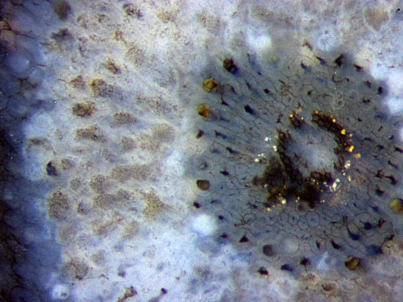
This image, as one may guess, has got much more to
offer than the appearance as a flower. It shows a cross-section from a
quite uncommonly preserved lower or subterranean part of Ventarura,
the last-discovered land plant in the Lower Devonian Rhynie chert. It
would not have been recognized as such if there were not lots of other Ventarura sections around. In fact,
there is no other plant species seen on the 6 cut faces of the 4 parts
into which this sample has been divided. Hence, one can safely assume
that
the section seen here is really Ventarura
although its state of preservation is so
peculiar that none of the other
about 500 sections seen on the cut
faces of this sample compares to this image. Nevertheless it may be
worth trying to
partially explain how the peculiar aspect is related to the structure
of the plant.
To begin with, the preservation in this case is such
that several concentric zones can be distinguished. The xylem in the
very
centre is decayed but a few of its small cells are still there,
arranged in a
small dark ring with gaps. The eye is attracted to the tiny bright dots
which greatly contribute to the illusion. They are
mineral precipitates formed around the xylem strand for reasons
unknown.
Next comes a broad ring which is usually
regarded as
phloem. It consists of cells with thin walls forming a clearly seen
network.
The dark dot or spot in the middle of every cell seems to be the
remains of the
more or less shrunken plasma.
The outermost cells of this ring are much
larger and
probably belong to the adjacent main part of the tissue called cortex.
Further
out in the cortex, the aspect is quite different. The former cellular
structure
can only be guessed from the pale grey spots with randomly distributed
dots like dust grains
possibly related to some fungus. Judging from experience, the
ubiquitous fungi
thriving
in the Lower Devonian habitat, as there are parasitic, symbiotic, and
saprophytic
ones, are
able to greatly modify the aspect of the living and dead plants before
they
become solid chert. Hence the effect of fungus activity has to be
considered in
this case, too. Some fungus known to invade a part of the
cortex of
other plants in the Rhynie chert,
appearing as a ring-shaped zone
on cross-sections, could be involved here, too. Still
farther out, the cortex tissue looks
more normal
again, with cell walls seen.
Note that this section does not show the ring
of well-preserved cortex
tissue which is characteristic for
the upper parts of this plant. Hence it can be assumed that this image
shows a section of a rhizome or of a lower part of a shoot. Perhaps
that ring is even more enigmatic
than the combination of effects contributing to this concentric
pattern.
H.-J.
Weiss 2016
 |
 |
96 |