Asteroxylon
tissue affected by fungi
 One may not expect this neat Asteroxylon
specimen in Fig.1, with the central xylem strand and two circles of
small strands branching thereof, to have been
in some
state of degradation while becoming silicified. Really, proceeding
degradation is indicated by the
absence of a clearly visible tissue structure. While other plants in
the Rhynie chert are usually seen in various states of preservation,
not seldom with very well preserved tissue, Asteroxylon is
never seen well preserved, except for the roots. Its early decay is
most probably due
to fungus activity. A few remains of the tissue are very faintly seen
here.
In idealised drawings, the
cross-section is occasionally shown
with large fungus-induced voids
arranged in a concentric ring as if
this were an intrinsic property of this plant, which it is not.
One may not expect this neat Asteroxylon
specimen in Fig.1, with the central xylem strand and two circles of
small strands branching thereof, to have been
in some
state of degradation while becoming silicified. Really, proceeding
degradation is indicated by the
absence of a clearly visible tissue structure. While other plants in
the Rhynie chert are usually seen in various states of preservation,
not seldom with very well preserved tissue, Asteroxylon is
never seen well preserved, except for the roots. Its early decay is
most probably due
to fungus activity. A few remains of the tissue are very faintly seen
here.
In idealised drawings, the
cross-section is occasionally shown
with large fungus-induced voids
arranged in a concentric ring as if
this were an intrinsic property of this plant, which it is not.
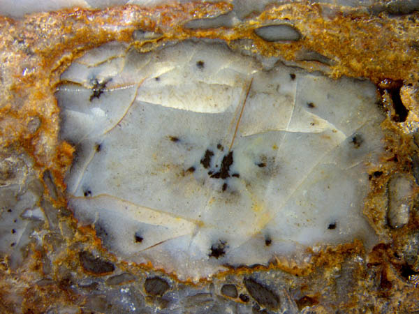
Fig.1 (right): Asteroxylon
cross-section with clear outline and xylem but faint remains of tissue.
Image width 11mm.
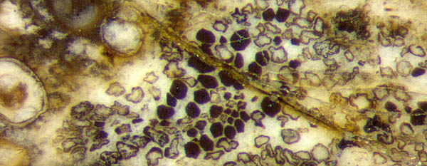
Fig.2 (left): Cross-section of disintegrated Asteroxylon xylem,
tracheids filled with dark fungus matter keeping their original shapes,
others shrivelled; fungus globules on the left.
Image width 1.4mm.
A fungus effect quite different from degradation is seen in
Fig.2, where a xylem strand has disintegrated into a bunch of separate
tracheids
for reasons unknown. Here, the tracheids are clearly subdivided into
two conspicuously different types: Those with black fills have kept
their
polygonal outline representing their original shape but the empty ones
have shrivelled. This phenomenon is seldom met with Asteroxylon.
A similar and likewise rare phenomenon is seen in Fig.3, where cells of
the soft
tissue are still visible because they had become filled with
fungus matter. The tissue between this uncommonly preserved area and
the central strand is seen to be degraded as usual, possibly due to
another fungus species. Notably there are no large voids
in Figs.1,3.
The dark fills are most probably made up of dense tangles of tiny
fungus hyphae which are not seen here. A hypha penetrating the wall of
a cell and growing a tangle inside has been found and documented
elsewhere [1]. 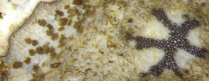
Fig.3: Asteroxylon
cross-section with central strand well preserved, adjacent tissue
heavily fungus-affected and degraded, outward area with some cells
filled with fungus matter, others hardly visible.
Image
width 4mm.
Cells filled with fungus matter, rarely seen with Asteroxylon, are a
common sight with Aglaophyton,
where the fungus is known to have thrived in the live plant [2] and has
got
the name Glomites
rhyniensis [3,9]. It is not
known whether this fungus had produced the cell fills in Figs.2,3. Asteroxylon had
probably been affected by more than one fungus, judging from
the phenomena and from the often abundant big resting spores
like those in Fig.2.
It is hard to believe that cell-size dark clots in more or less decayed
fossil
plants have repeatedly been misinterpreted as mite coprolites, despite
of glaring contrary evidence: angular shapes fitting to the cell
cross-sections [4], sizes fitting to the tissues of bigger or smaller
cells [5], often
arranged in rows fitting to
the rows of
cells [2,6], and finally the absence of any fossil mites in the samples
with the alleged coprolites. Apparently the idea of mite coprolites had
become self-accelerating, with feedback provided by mutual
encouragement within the community of coprolite fans. Persistent
warning against the
obviously nonsensical coprolite idea [7] has
hopefully ended that craze in 2016. The fungus clots in
Asteroxylon (Fig.3)
had been part of this effort since 2009 [8], and the recently found
clots in Fig.2 may add to the abundantly available evidence against
angular clots being coprolites.
The proponents of the
once favourite mite coprolite hypothesis apparently do not mention it
any more but
do not retract it. Hence, it is still lingering in the palaeobotany
literature, spreading confusion. Therefore it should be contradicted
anew with every new evidence, which has been done here.
H.-J.
Weiss
2018
[1] H.
Kerp:
De Onder-Devonische Rhynie Chert ... . Grondboor& Hamer
58(2004),
33-50. See
image here: Fossil Wood
News 4,
Fig,3.
[2] Rhynie Chert News 85.
[3] T.N. Taylor
et al.: Fossil arbuscular mycorrhizae from the Early
Devonian. Mycologia 87(1995), 560-73.
[4] Fossil Wood News 5 , Figs.1-5; 8,
Figs.4,5; 18,
Figs.1-5.
[5] Fossil Wood News 18, Figs.1,2.
[6] Fossil wood News 8, Figs. 2,3,9;
[7] Google: coprolites Roessler
[8] Rhynie Chert News 28,
[9] T.N.Taylor, M. Krings, E.L. Taylor:
Fossil Fungi, Elsevier 2015, p121.
 |
 |
124 |