Croftalania
images
Picturesque fossil cyanobacteria are seldom
seen. The Rhynie
chert, which is famous for its often well preserved Lower Devonian
organisms, offers such, too. The filamentous cyanobacterium Croftalania [1],
when
grown in dense bunches on substrates, can make fanciful appearances [2 ,3].
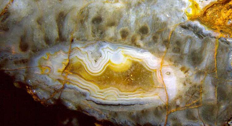
Fig.1: Croftalania
tufts on agate in Rhynie chert.
Width of the picture 8.6mm.
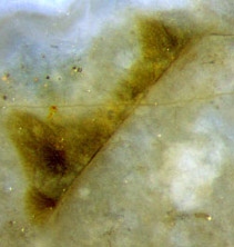
Figs.2 and 3 (smaller one below): Croftalania
tufts on plant cuticle.
Width of the pictures 4.3mm,
0.9mm.
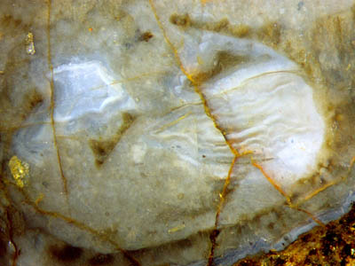
Although Fig.2 is less conspicuous than Fig.1,
it is more instructive. In Fig.1 it is not quite obvious on which
substrate Croftalania had
grown because the agate had formed later in the water-filled cavity.
Inspection of Fig.2 suggests that in Fig.1, too, it had grown
on
the cuticle of a decayed land plant, probably Aglaophyton,
of which only a roughly elliptical shape is left. The instructive
feature in this picture is the thin line detaching from the periphery
in the upper half on the right and dangling loosely inside. It is the
cuticle which had served as the substrate for Croftalania
but is pried loose from it now. (One
should ignore the stained cracks which do not provide any information
here.)
This
configuration permits some conclusions concerning the mechanical
properties of the components. The coating consisting of Croftalania
has preserved the contour of the vanished plant
tissue while the cuticle
was peeled off, hence
it must have been stiff. Such stiffness is compatible with the
assumption that the cyanobacteria filaments were embedded in organic
gel and that all tufts of the coating were fused into a continuous
layer of gel. Patches of the layer broke off, one is now seen still
adhering to the end of the detached cuticle, enlarged in Fig.3, where
the (displaced) cuticle is clearly seen as a thin dark line. If no gel
were
involved
and the filaments were simply dangling in the water, the tufts sticking
to the end of the detached cuticle would probably have become deformed
by
the motion. The absence of
any
visible deformation is another argument for the presence of gel between
the filaments.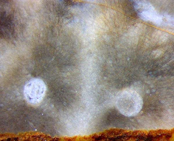
Often the tufts show a textured appearance, as expected, owing
to the directed growth of the filaments. The growth is usually
more vigorous upwards to the sunlight, as seen in Fig.1 and in several
pictures of [2].
In
the present sample the texture due to growth is often superimposed by
phenomena which might be degrading effects in the organic gel between
and around the filaments. The radial growth direction with respect to
the cylindrical cavities in Fig.4, still faintly seen here,
has
become
overlaid with equally faint lines of other orientation and with small
whitish spots.
Fig.4 (right): Croftalania
grown on unidentified plant parts. The radial arrangement of the
filaments is apparently spoilt by secondary phenomena in the gel. Width
of the picture 2mm.
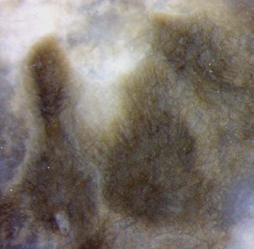 Fig.5
(left): Croftalania
tufts influenced by shaping after growth and by the formation of a
network of narrow cracks of uncertain cause.
Fig.5
(left): Croftalania
tufts influenced by shaping after growth and by the formation of a
network of narrow cracks of uncertain cause.
Width of the picture 1.4mm.
In the absence of apparent degradation, as in the sample described in
[2], silicification turned both the organic
gel and the surrounding water into
clear chalcedony. The phenomenon of clear substances turning white is
always due to the formation of grains or holes with sizes comparable to
the wavelength of light. Hence, the patches of whitish chalcedony
in the present sample could have been brought about in different ways:
The organic gel itself could have become whitish by degradation, or
substances released in degradation could have triggered crystal growth
during silicification such that the chalcedony became whitish. By the
way, whitish chalcedony improves the visibility of the cyanobacteria
filaments lying near the surface of the chert.
The
club-shaped tufts in Figs.1,5 indicate the action of processes other
than mere growth of filaments. With the presently available fossil
material and facilities, only a minor part of the arising questions can
be answered. The clear-cut outlines of the tufts which do not
correspond to the bunch of aligned filaments, as seen on parts
of
Fig.5 and much more distinctly in [2],
had most probably been shaped by grazing aquatic creatures, an idea
supported by the presence of the crustacean Castracollis on the
"blue-green meadow".
All pictures have been taken from the cut faces of one
chert sample of 0.19 kg obtained in 2014.
H.-J.
Weiss
2015
[1] M. Krings, H. Kerp, H. Hass, T.N.
Taylor, N.
Dotzler:
A filamentous
cyanobacterium showing structured colonial growth from the Early
Devonian Rhynie chert.
Rev. Palaeobot. Palyn.
146(2007), 265-276.
[2] H.-J. Weiss:
Croftalania venusta
and other Lower Devonian microbes. Rhynie Chert
News 56.
[3] H.-J.
Weiss: Rhynie chert -
Implications of new finds. European Palaeobotany and
Palynology Conference 2014, Padua.
 |
 |
72 |