Horneophyton
detail
Several contributions on Horneophyton
in the scientific literature
concern sporangia [1,2,3], epidermis [4], and the tubers in the ground
[5]. Additional evidence concerning the often odd-shaped sporangia,
the
peculiar preference of the tubers to form level
bands inside,
and the arrangement
of the tubers in strings has been
presented on this website.
Apparently
not much attention has been paid to the tuber tissue because most often
there are no distinctly seen details except for the rugged dark base of
the
central strand (Fig.1). The present specimen is uncommon with an
unexpected boundary
inside the tuber, marked by more clearly seen cells with
whitish lumina and dark stains near the walls. As seen in
Fig.2, this coarse boundary separates
tissues of different structure.
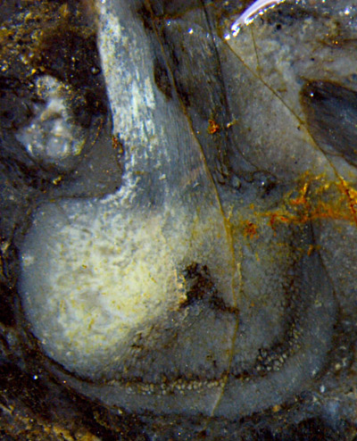
Fig.1: Horneophyton
tuber, 5mm wide, on the raw surface of a Rhynie chert
sample. Note the uncommon boundary above the bottom. Rhizoids
and part of the central
strand are missing here.
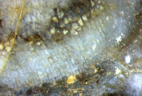 Fig.2: Horneophyton
tuber, detail from Fig.1 below right. Note the texture of
the tissue between the dark boundary and the bottom. Width 1.7mm.
Fig.2: Horneophyton
tuber, detail from Fig.1 below right. Note the texture of
the tissue between the dark boundary and the bottom. Width 1.7mm.
Above the boundary the cells are polyhedral without an apparent order.
Below the boundary the cells are smaller and tend
to be rectangular and arranged in files directed towards the
bottom.
The question arises whether or not this particular structure formed by
the exceptional tuber in Fig.1 is also found, possibly less
conspicuous, in other specimens. In Fig.3,
taken from the same chert sample only about 1cm away from
the big tuber in Fig.1, some of the lowermost cells are nearly
rectangular and elongated, oriented towards the bottom, perhaps
in connection with the rhizoids grown from there. A similar texture of
part of the tissue is more distinctly seen in rhizomes of other plants
in the Rhynie chert. In the case of Horneophyton,
this easily escapes notice because the
tissue
of the tubers is most often degraded and not seen as clearly as in
these images. Also there are tubers whose lowermost cells do not seem
to differ from the others.
The question whether textured tissue had always been present at the
bottom of the tubers, even if not well seen after degradation
and
silicification, may be answered by means of additional fossil
evidence.
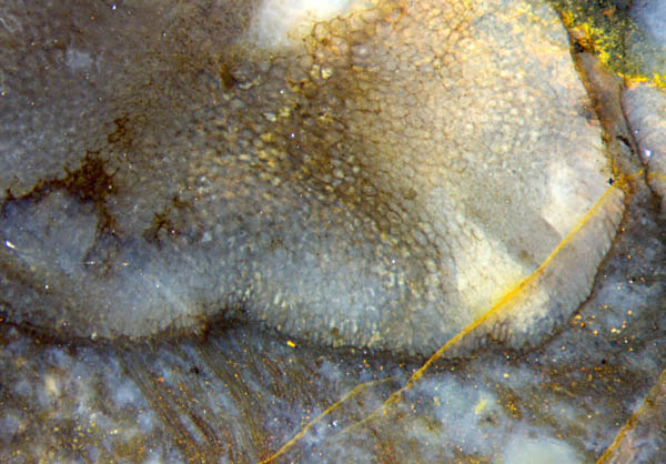
Fig.3: Horneophyton
tuber with rhizoids on a cut face of the same sample as in
Fig.1. Width of
the picture 4.3mm
H.-J. Weiss 2015
[1] A.A. Bhutta:
Observations on the sporangia of Horneophyton
... , Pak. J. Bot. 4(1972), 27-34.
[2] D.A. Eggert:
The sporangium of Horneophyton
... ,
Amer.J. Bot. 61(1974), 405-413.
[3] W.El-S.
El-Saadawy, W. Lacay: The
sporangia of Horneophyton
...
, Rev. Pal. Palyn. 28(1979), 137-144.
[4] H.
Hass: The epidermis of Horneophyton
... , N. Jb. Geolog. Paläontolog. Abh. 183(1991),
61-85.
[5] H.
Hass, H. Kerp:
Rhynie chert plants and their substrates. "The
Rhynie Hot Spring System", Aberdeen 2003.
 |
 |
81 |