Nematoplexus
aspects
The nematophyte
Nematoplexus,
discovered by
A.G. Lyon
[1] in one piece of Rhynie chert and since then found in poorer quality
only a very few times more, seemed
to be characterized by rather well defined structure parameters, as
pointed out
in Rhynie
Chert News 29:
(1) The tubes are wound into a rather
regular screw-like
thread ...
(2) ... which is always right-handed ...
(3) ... with diameters of 0.08 - 0.12 mm.
(4) ... whose pitch seems to roughly equal its diameter.
A sample found in 2003 confirmed this, except for the diameters of
tubes and threads:
(3) ... with thread diameters of 0.14 - 0.21 mm.
Closer inspection of the raw surface of this
sample has revealed another patch of
3mm width with the usual curled tubes and, surprisingly, a few tube
sections curved and wound so slightly that it is uncertain whether
they are parts of bigger threads, wound otherwise, or flat (Fig.1,
left).
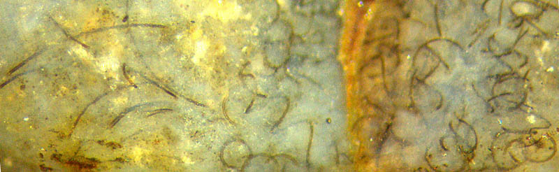 Fig.1:
Nematoplexus,
thread-like tubes and weakly curved ones. Width
2.6mm.
Fig.1:
Nematoplexus,
thread-like tubes and weakly curved ones. Width
2.6mm.
Fig.2: Detail of Fig.1. Width 0.62mm.

Apparently they are rather evenly curved but their radius of curvature
is difficult to estimate owing to the uneven surface of the raw
sample and limited visual depth. Some of the slightly
curved tubes are near an edge of the sample so that by visual
inspection from various sides an osculating plane to the tube fragment
can
be defined. By turning the sample such that the osculating plane is
made the picture plane of the photograph, the radius of curvature
is easily found as 0.75mm for one
of the tube fragments. It would not much differ from the half diameter
of the thread (if this tube were wound, which cannot be ascertained
with the available fragment).
So it appears that Nematoplexus
offers more enigmas: In addition to the small regular threads, it
brings forth evenly curved objects with diameters
nearly ten times larger but made
of tubes of about the same
thickness of 10...15µm, also slighty curved thinner tubes of 7-8µm
(Fig.2, two thin tubes running parallel with
only a small gap in between).
This contribution has shown structures which distinguish Nematoplexus from
other nematophytes.
The following is about one feature which Nematoplexus
has in common with other nematophytes: clots called
"branch-knots" after [1]. Since nobody seems to know what nematophytes
are, one need not wonder why nobody knows what the branch-knots are
for. They look as if they produce the tubes. Other
silicified nematophyte samples seem to indicate that the branch-knots
also contain
a tangle of tubes which
are much thinner than the conspicuous larger ones seen here. (See
Annotation 2020.)
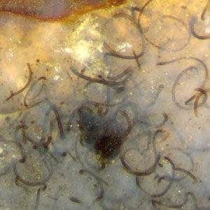
Fig.3: Nematoplexus
branch-knot amidst spiralling tubes. 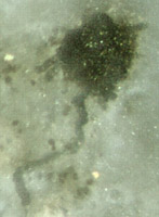 Width of the picture 1.6mm
1mm.
Width of the picture 1.6mm
1mm.
Fig.4: Solitary branch-knot of
Nematoplexus
with short tubes attached but no spirals nearby. Same chert sample.
Width of the picture 0.25mm.
The solitary knot of about 0.12mm in Fig.4 resembles the one pictured
in
[2] under "another branch knot" (as the images are not numbered there),
with the difference that the diameter of the tubes is less than half
that in [2]. The
similarity of the pictures (except sizes) of solitary knots here and in
[2]
suggests that they are no incidental formations but represent
another type of knot or another stage of development without the
spiralling tubes as in Fig.3.
It appears that Nematoplexus
is a subject more difficult to handle than originally thought, for
quite different reasons:
-
The sample containing the type specimen first described in [1] had been
shattered by A.G. Lyon
unawares
of the unique content of the sample, with a
hammer, and only some fragments had been recovered.
(Fossiliferous chert should never be hit.)
- Tube sizes may differ
distinctly:
- The smooth tubes of one specimen may differ in their radius
of curvature by a factor of about ten or more: Fig.1.
- The available information is scarce, published size data
are
contradictory or questionable: [3] differs from [1,2].
In particular, the
scale bar of Fig. 6.9 in [3] should be 18µm instead of 100µm, which
follows from comparison
with [2]. What is offered as a branch knot in [3], Fig.
6.10, differs much from any Nematoplexus
branch knot pictured
here and in [1,2], which raises the suspicion that a wrong picture has
been placed in [3] by mistake. The suspicion is confirmed by
the
presence of several more mistakes and errors concerning Rhynie Chert
fossils in [3], Chapter 8.
Considering that the rugged outline of
the branch knots is poorly defined, it is quite unreasonable to
quantify a lower size boundary as 99µm, as done in [2].
The characteristic feature of Nematoplexus,
the
spirally wound tubes, is ascribed in [2] to all nematophytes:
"Nematophytes appear to generally comprise networks of intertwined
spirally coiled tubular cells."
This is utterly wrong: No other nematophyte has got spirally wound
tubes.
According to present knowledge, Nematoplexus
is very rare but less rare in mm-size patches than in larger
aggregates. Hence
there is reason for hope that more specimens
will be discovered by carefully inspecting the Rhynie
chert samples stored in collections.
All pictures have been taken from the raw surface
of a chert sample of 0.28kg found
in 2003, labelled Rh9/86.
Annotation
2018: (A scale error in Fig.3 has been
corrected.) For more information on Nematoplexus
see Rhynie
Chert News 122.
Annotation 2020: For more information on Nematophytes
see Rhynie
Chert News 156.
H.-J. Weiss 2013,
2014,
2015, 2018, 2020
[1]
A.G. Lyon:
On the
fragmentary remains of an organism referable to the nematophytales,
from the Rhynie
chert, Nematoplexus
rhyniensis.
Trans.
Roy. Soc. Edinburgh 65(1961-62), 79-87, 2
plates. (Scale
error on Plate I Fig.1: not x19 but x1.5)
[2]
www.abdn.ac.uk/rhynie/nemato.htm
[3] T.N. Taylor,
E.L. Taylor, M. Krings: Paleobotany,
Elsevier 2009.
 |
 |
51 |