A rare sight of Ventarura
tissues (2)
 As already stated earlier
under this heading (Part 1), Ventarura
is
distinguished by a conspicuously differential persistence of tissues
before silicification of the dead plant. A cylindrical tube, seen on
cross-sections as a concentric ring of well-preserved
cells amidst decayed tissue, is always an indication of the presence of
Ventarura
in the chert sample. According to the palaeobotany literature the epidermis of Ventarura has never been seen [1,2]. For whichever
reason, it had been preserved seldom. Meanwhile, it has been seen only
twice when inspecting the surfaces and cut faces of about two dozen own
chert
samples with Ventarura.
One is shown in Part 1, the other one in this contribution
(Fig.1).
As already stated earlier
under this heading (Part 1), Ventarura
is
distinguished by a conspicuously differential persistence of tissues
before silicification of the dead plant. A cylindrical tube, seen on
cross-sections as a concentric ring of well-preserved
cells amidst decayed tissue, is always an indication of the presence of
Ventarura
in the chert sample. According to the palaeobotany literature the epidermis of Ventarura has never been seen [1,2]. For whichever
reason, it had been preserved seldom. Meanwhile, it has been seen only
twice when inspecting the surfaces and cut faces of about two dozen own
chert
samples with Ventarura.
One is shown in Part 1, the other one in this contribution
(Fig.1).
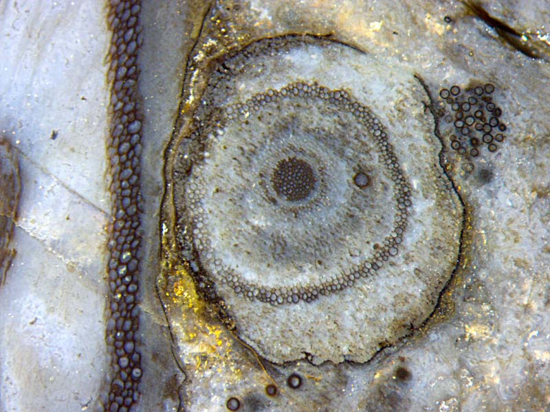
Fig.1: Two sections of Ventarura
with strongly differing
aspect:
- inclined section, partially seen on the left, tissues
decayed except xylem and persistent tube,
- cross-section, cortex decayed, patches of epidermis
clearly preserved,
black stained coating on most of the cuticle and
on cell walls (except epidermis),
and on fungus spheres.
Width of the picture 5.5mm.
Incidentally,
Fig.1 does not only show the epidermis along more than half the
circumference of the cross-section. It also gives an impression of the
variable aspect of the silicified plant. Partially seen on
the left is an inclined cut of a shoot, width 5mm. The part of
the
elliptical section is nearly straight here due
to deformation by
contact with the shoot seen in cross-section. The latter is also
deformed. Its original diameter appears to have been 3.3mm. Although it
is not much smaller than the left one, the cells of its persistent ring
are much smaller. The black coating is thin on
the cell walls. No black coating is seen on the left side of the
persistent ring and on the epidermis cell walls (Fig.2). The black
coating is thick on the cuticle, on the
nearby fungus spheres, and on the walls of the big cells of the shoot
on the left. A
black coating is also on the xylem cell walls and on phloem cells
loosely arranged as a ring around the xylem. A few scattered
dark dots
are of a quite different nature, they are cells filled with dark fungus
matter.
The
cortex has been subdivided into inner cortex, mid-cortex, outer cortex
[1,2].
Apparently this subdivision is not meant to explain anything. It is
only meant to assign names to the areas of the cross-section separated
by
the well-preserved ring called
mid-cortex for this purpose.
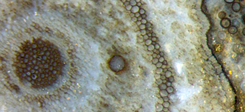
Fig.2 (below right): Components of partially
degraded Ventarura
cross-section:
central xylem strand, phloem (partially with stained cell walls),
decayed inner cortex with fungus hyphae, well preserved mid-cortex with
cell walls coated and stained dark, decayed outer cortex, epidermis,
cuticle coated and stained dark; fungus chlamydospores with and without
dark coating.
Width of the picture 2.2mm.
In
particular, the mid-cortex is not a separate tissue with definite
boundaries since on either side of the mid-cortex there are always
cells which do not belong to it as a whole but only with part of their
walls. How this is brought about, and for what purpose, is still
enigmatic.
Lots of fungus hyphae, or the remains thereof, are seen randomly
distributed, together with debris from the tissue, in the
areas called the inner and the outer cortex. Traces of the former
cortex tissue structure are still vaguely seen below the epidermis on
the right.
The
thickness of the epidermis is rather uniform, about 70µm. The width of
the elongated epidermis
cells aligned along the shoot (see
Rhynie
Chert News 61,
Figs.5,6) can appear
variable, depending on their position with respect to the cut plane.
In this sample the seldom preserved epidermis extends across the
cut gap into the other slab but has not been traced farther.
H.-J.
Weiss
2016
[1] C.L.
Powell, D.
Edwards, N.H. Trewin: A new vascular plant from the
Lower Devonian Windyfield chert, Rhynie, NE Scotland.
Trans. Roy. Soc. Edinburgh, Earth Sci.
90(2000 for 1999), 331-349.
[2] D.
Edwards: Embryophytic sporophytes in the Rhynie
and Windyfield cherts.
Trans. Roy. Soc. Edinburgh,
Earth Sci.
94(2004 for 2003), 397-410.
 |
 |
91 |