Microbial debris in Rhynie chert
Black dust grains or flakes of no definite shape, and loose
aggregates thereof, are not rare in the Rhynie chert. They may be found
among tissue or along former gel surfaces formed during silicification
of the silica-rich water. The shapeless black
debris is seen in Fig.1
below the fungus sphere, in the narrow gap of gel cracks, and along the
faces of wide gel cracks filled with bluish chalzedony.
Observations on several samples have raised
the suspicion that the black matter is possibly the carbonaceous
remains of degraded
microbial coatings. The present sample offers evidence in favour of
this idea.
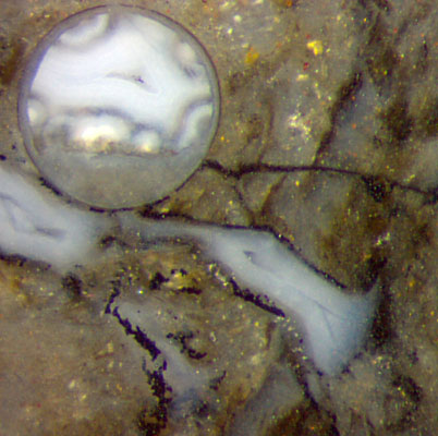
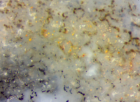
Fig.1 (far left): Small amounts of black matter along former silica gel
surfaces.
Fig.2 (left): "Hard" black debris below and "soft" brown formations
above, suggesting microbial colonies on former silica gel surfaces not
seen here.
Height of either picture 1mm.
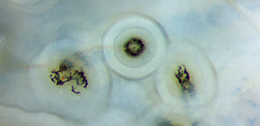
Fig.3 (below): Hyphae of an aquatic fungus
coated with silica gel, overgrown with microbial slime turned into
black debris, finally coated with more layers of silica
gel.
Width of the picture 1.3mm, same magnification as Figs.1,2.
The brown formations of fluffy appearance as seen in Fig.2
are seldom
preserved in
the chert. Probably they degraded and shrunk into
black debris soon. This is supported by arrangements like the middle
one in
Fig.3, where a microbial colony grew on a coated hypha of
an aquatic fungus inside hollow Aglaophyton
(not shown here),
then
decayed
soon and turned into black debris before further silica was being
deposited. Deposition went on for a longer while until a thick
multi-layered
coating, diameter 0.34mm, had built up. The less compact arrangement of
the debris around the hypha on the left in Fig.3 seems to be due to
branching of the hypha. It shows that the debris is
essentially the same as in Fig.2.
What
is seen on the present sample seems to be related to other
phenomena in the Rhynie chert. As mentioned earlier, the dark aspect
usually observed with hollow straws of Aglaophyton and
with the dark
rings on cross-sections of Ventarura
is probably brought about by microbial deposits on
the cell walls, and "the dark deposit is not
always strongly bonded to the substrate but may become reduced to
detached flakes", as stated in Rhynie
Chert News 83
and shown in Rhynie
Chert News 60,
Fig.7.
In view of the related observations mentioned here it may be
justified to say that Fig.2 offers a
rare sight strongly suggesting an interpretation of
the black
dust-like particles as the remains of
microbial colonies.
H.-J. Weiss
2015
 |
 |
87 |