Comment on: The Late Palaeozoic tree
fern Psaronius ... by R. Rössler
[1]
The subject of that paper, the intimate connection of Permian tree
ferns with the ecosystem, is doubtless an interesting one, and thus the
paper, too, had been destined to become interesting. The comment does
not concern this aspect of the matter but the tangle of minor
inconsistencies due to superficiality and the more serious indications
of questionable judgement, unseen at first sight but emerging clearly
with closer inspection, having escaped the notice of the reviewers and
numerous palaeobotanists whose "critical and stimulating discussions"
are
acknowledged in the paper.
From the aspect of the Ankyropteris
cross-sections in Plate VII one can
already guess that the magnification data are mutually incompatible.
With some effort one can find data which hopefully will lead
near the real sizes. One
can make use of the fact that the same pictures have been published by R. Rössler
several
times,
with variation in scale, orientation, frame boundary, and also as
mirror images. As an additional difficulty, the modified images, too,
come with contradictory size data, which delays but does not prevent
comparison. 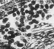
Fig.1: Angular clots in the tissue of the climbing fern Ankyropteris
brongniartii, detail from [1], Plate VII4,
Lower Permian,
Chemnitz.
Provided that one can trust the magnification given as 6x for
Fig.334 in [2], one
can conclude from Fig.336, which is the mirror image of a detail of
Fig.334 and
is also the same as Plate VII5 in [1] turned
around by 135°, that 35x
instead
of 14x is the proper magnification number for Plate VII5,
after
correcting the
scale of Fig.336 by a factor of 2 which is evident from comparison with
Fig.334.
Minor size differences up to 30% become apparent by comparing the
figures on
Plates III, IV, VI in [1] with those in [2] and [3].
Fig.336 in [2] is said to show a detail from
the main axis of Ankyropteris
in Fig.334. There is no such detail on the main axis. The detail is
found on
the frond stalk. The same error is present in [1], caption to Plate
VII5.
(Here, the meaning of
"Ankyropteris marginal axis" is not immediately
obvious but
from Fig.334 in [2] it can be deduced that it means a main
axis of the
climbing fern which is not completely enclosed within the Psaronius
trunk.)
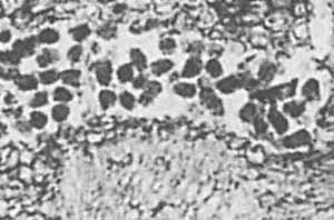
Fig 2: Cell-size angular clots in Ankyropteris, arranged
in
files on the left, detail from [1], Plate VII2.
Plate VII3-5 shows details of a thin slab
pictured in a paper by Sterzel
[4],
who was careful enough to
refrain from an interpretation of the tiny dark clots seen in these
pictures. Along with such clots in Callistophyton
(Plate VI), they
are interpreted in [1] as arthropod
coprolites: "Two distinct
size
orders of the coprolites indicate coexistence of different arthropods
or different ontogenetic stages ..."
The interpretation of these and other cell-size
clots is specified
as oribatid mite coprolites in [2,3].
Fig.3 (right): Angular clots of various sizes
in Ankyropteris, detail
from [2], Fig.335 below right, (which is the mirror image of Plate VII4
in [1]).
Note the
cell above right with a clot of corresponding shape inside.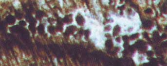
The publications [1-3] are obviously influenced
by the notion of
Palaeozoic oribatid mite coprolites which had spread among
palaeobotanists in
the 1990s, apparently by adopting without checking, even
though no oribatid mite had been spotted in all Carboniferous, Permian,
and
Triassic [5]. Contrary evidence, although rather conspicuous, was not
noticed
or ignored: Clots with polygonal outline indicating angular shape
(Figs.1-3)
compatible with the sizes and shapes of nearby cells
(Figs.2,3), occasionally arranged in files compatible with the cell
files of
the surrounding tissue (Fig.2), also as separate clots inside cells
(Fig.3).
There are different types of tissue with
differential cell sizes in the Ankyropteris
axis. The clots in Fig.2 are compatible with the cell lumina of the
tissue. Two
chains of clots on the left look as if they had not been dropped there
randomly. They look as if they had kept their positions where they had
formed
within the cells, a few remaining of which are seen below left. This
suggests
that the clots are casts of the cell lumina left over after the cell
walls of
the tissue which had been there where the clots are now had decayed.
This idea
is supported by several more observations based on
own samples and
pictures in other publications. 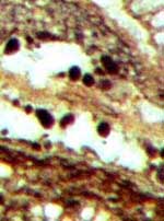
Fig.4: Clots of various shapes and sizes in Ankyropteris,
detail
from [2], Fig.430, which is the same picture, upside down, as Plate
VII1 in [1].
By looking carefully at the pictures in [1,2,3]
one finds abundant
evidence contradictung the claim that there are "two distinct size
orders" of "coprolites". There are any sizes
between tiny and rather big (Fig.4), like the cell sizes in the
vicinity. Also the
arrangement of these clots suggests that they had not been randomly
dropped
into a cavity by some creature.
The observation that the variation of
sizes and
shapes of the clots
is the same as with the cells of nearby tissues provides another
argument
against the coprolite interpretation.
Finally it can be stated that lack of thoroughness has led to both
the contradictory size data in [1,2,3] and the misconception of
cell-size
coprolites,
which is still being upheld [6] despite of repeated warnings [7,8].
Wood
rot
due to fungus or microbial activity has to be taken into consideration
as a cause of clot formation.
Samples: Museum für Naturkunde Chemnitz.
Annotation 2017:
After several more publications trying to make believe that mite
coprolites in fossil wood are real, the latest one from 2015 [9], and
after the presentation of lots of contrary evidence on this website, Rössler has
apparently given up the idea.
H.-J. Weiss 2011,
2017
[1] R.
Rössler : The late palaeozoic
tree fern
Psaronius - an ecosystem unto itself.
Rev. Palaeobot.
Palyn. 108(2000), 55-74.
[2] R.
Rößler : Der versteinerte Wald von Chemnitz, 2001.
[3] R.
Rössler : Between precious inheritance and immediate
experience.
in: U. Dernbach, W.D. Tidwell
: Secrets
of Petrified Plants. D'ORO Publ., 2002, 104-119.
[4] J.T. Sterzel:
Die organischen Reste des
Kulms und
des Rotliegenden der Gegend von Chemnitz.
Abh. Königl. Sächs. Ges. Wiss.,
Math.-phys. Kl. 35(1918), 205-315.
[5] C.C.
Labandeira,
T.L. Phillips, R.A. Norton : Oribatid
mites and the decomposition of plant tissues in paleozoic coal swamp
forests.
Palaios 12(1997), 319-53.
[6] M.
Barthel, M. Krings, R. Rößler: Die
schwarzen Psaronien
von Manebach, ihre Epiphyten, Parasiten
und Pilze.
Semana*
25(2010), 41-60.
*( recently re-named, former name: Veröff.
Naturhist. Mus. Schleusingen)
[7]
H.-J. Weiss:
Rätselhaftes aus Hornstein
und Kieselholz.
6. Chert Workshop 2007,
Naturkunde-Museum Chemnitz.
[8]
H.-J. Weiss:
Märchenhaftes und
Ernsthaftes im Hornstein. 8. Chert Workshop 2009,
Naturkunde-Museum Chemnitz.
[9] Z. Feng, J.W. Schneider, C.C. Labandeira, R.
Kretzschmar, R. Rössler: A specialized feeding habit of Early
Permian oribatid
mites.
Palaeogeography,
Palaeoclimatology,
Palaeoecology 417(2015), 121-124.
|

|
 12 12 |