Enigmatic
knots of an enigmatic
organism
 The wound tubes of the
twice enigmatic Nematoplexus
[1] mentioned in [2] under the heading Enigmatic
Organisms are supposed to be produced
inside "branch knots" although branching
apparently has never been seen there.
What is offered as a branch knot in [2],
Fig.6.10, looks rather like a fluid structure governed by surface
tension on a substrate with variable
wettability, thus being in no way related to the branch knots shown
here.
The wound tubes of the
twice enigmatic Nematoplexus
[1] mentioned in [2] under the heading Enigmatic
Organisms are supposed to be produced
inside "branch knots" although branching
apparently has never been seen there.
What is offered as a branch knot in [2],
Fig.6.10, looks rather like a fluid structure governed by surface
tension on a substrate with variable
wettability, thus being in no way related to the branch knots shown
here.
Branch knots of a hitherto unknown type with big tubes have recently
been discovered (see Rhynie
Chert News 71
and Figs.5,7,9 below right).
They are quite surprising for their features:
- tube diameters >20µm, 30µm at the base,
- tubes nearly straight or slightly curved,
- found only in samples with the usual Nematoplexus
spirals.
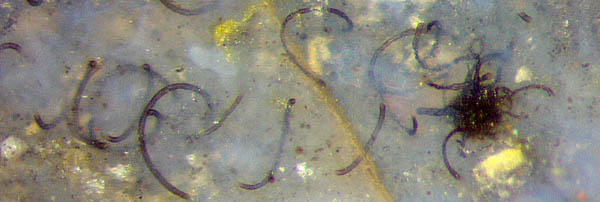
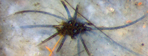
Most often, knots are found among scattered spirals
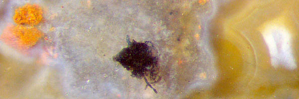
(Figs.1,2). Less often, solitary
knots of various aspect are found in areas where regular spirals
are
absent (Figs.3-9) but usually not far away in the chert sample. (Note
that "spiral" is meant here short for "wound in
a screw-like way".)
The solitary knots add to the mystery surrounding Nematoplexus.
Attached to these knots are tubes
differing in shape and size from the "normal" screw-like ones with
their
remarkably constant curvature and twist, for reasons unknown.
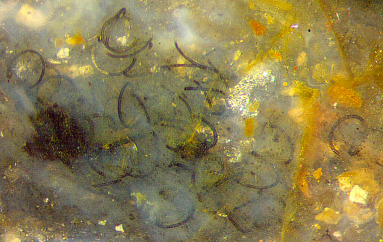
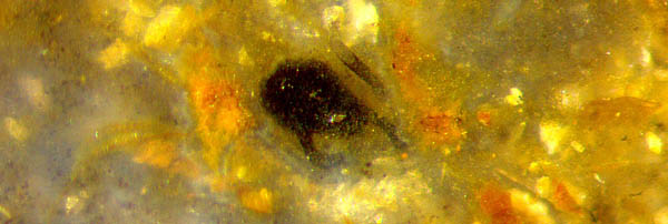
Fig.1:
Nematoplexus
spirals and parts thereof seen in pale bluish chalcedony;
big dark "branch knot", small one near the middle of the image. Note
that there is a spiral with three turns above the knot on the left and
another one right of the middle, with one tangent of the spiral
incidentally being perpendicular to the picture plane so that the
spiral looks like an inclined "3". Width of the picture 1.73mm.
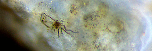
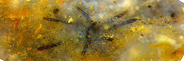
Figs.2-9: "Branch knots" of
various aspect, with or without spiralling tubes nearby. Same scale as
Fig.1, width of the pictures 1.38mm.
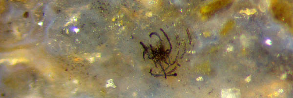
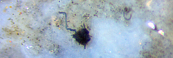
With the circular cross-sections of the cut-off big tubes in
the last image clearly seen, the question arises
what the onsets of
the tubes inside the knot might
look like. Are they all interconnected or did
they grow from separate points ? Apparently there is no answer
to simple questions of this kind at present. *
Non-spiralling tubes may show annular
or spiral wall patterns.
Dozens of tubes with patterned wall have been found loosely arranged in
a lump resembling Nematothallus
(see Rhynie
Chert News 107),
only millimeters away from the typical Nematoplexus
spirals
but in no way connected to the latter. The suggestion that
Nematoplexus
"may represent the permineralized equivalent of Nematothallus" [2]
is not at all substantiated.
As another extension of the Nematoplexus
enigma, the regularly wound tubes, too, are occasionally
seen with distinctly differing sizes, even at the very same spot
of the sample (Rhynie
Chert News 106,
there Fig.2). Also it appears that tubes with
diameters smaller than
those of the regular spirals can be weakly curved or nearly straight,
like the one seen crossing the last image here. Furthermore, very thin
slightly curved tubes (4µm ?) apparently come out of the big black
knot.
One may wonder how the various details thought
to be parts of Nematoplexus
can possibly be mutually compatible so that they can be regarded as
aspects of one species. Otherwise they might represent different
species grown very near to each other in places attracting
nematophytes. One might even suspect that different nematophytes could
have more or less united to form a kind of symbiosis.
In view of the variety of structural features associated with Nematoplexus, it
can
be expected that this nematophyte will yield more surprises
with more finds turning up in Rhynie chert samples stored in
collections.
* Annotation 2020: Apparently the tubes seen outside the "knots" had not branched inside.
Samples from own collection:
Rh6/102 (0.03kg) found by
S.
Weiss in 2003: Figs.1,5,7;
Rh9/86 (0.28kg) own find in 2003, Part 2: Figs.2,4,6,8 (left
column of images);
Rh15/79 (0.27kg) obtained from Barron
in 2014, Part 1: Fig.9, Part 2: Fig.3.
H.-J.
Weiss
2018 (2nd
version) 2020
[1] A.G.
Lyon:
On the fragmentary remains of an organism referable to the
nematophytales, from the Rhynie chert, Nematoplexus rhyniensis.
Trans. Roy. Soc. Edinburgh 65(1961-62), 79-87, 2
plates.
(Scale error on Plate
I Fig.1: not x19 but x1.5)
[2] T.N. Taylor,
E.L.Taylor, M. Krings: Paleobotany, Elsevier 2009. (Scale error on Fig.6.9: scale bar not 100µm, rather 20µm ?)
 |
 |
126 |