Enigmatic black deposit in Rhynie chert
There are dark or black deposits of various
type in the Rhynie chert: at the bottom of once water-filled pockets
present
during silicification, as level plates in such pockets or within Horneophyton
tubers, on detached
or adhering plant cuticles, or on cell walls of
early land plants. In the case of Ventarura,
cells with
dark deposit on the walls have been interpreted as sclerenchyma, which
is most probably erroneous [1]. The black deposit shown here is of a
different aspect. It looks as if once it had been an oily substance
within a narrow gap of a crack, then had become solidified and
glossy.
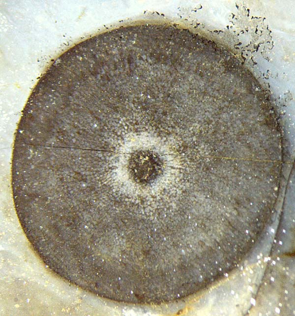
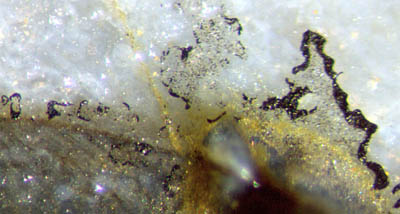
 Figs.1-3: Aglaophyton
cross-section, 4 mm, with dotted areas along the
circumference, partially bounded by glossy black blobs
of various shape; image successively enlarged.
Figs.1-3: Aglaophyton
cross-section, 4 mm, with dotted areas along the
circumference, partially bounded by glossy black blobs
of various shape; image successively enlarged.
As seen on the Rhynie chert fragment in
Figs.1-5, glossy black blobs are irregularly arranged near the
well-preserved cross-sections of Aglaophyton.
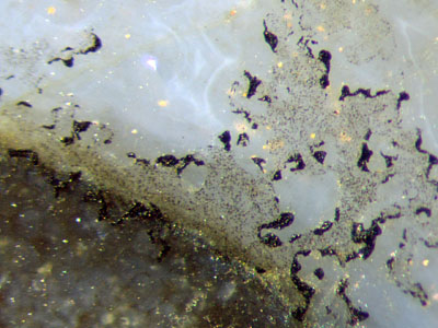
Remarkably, the blobs are seen ...
(1) only on the matt upper fracture face of the
chert
layer fragment,
(2) nearly always on the very face but not inside
the chert,
(3) always at or not far from either side of the
plant contour,
(4) bounding dotted areas.
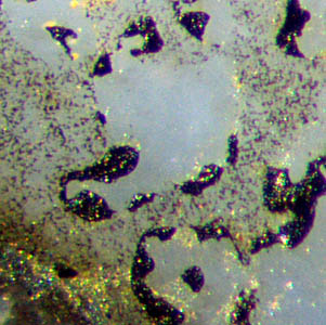
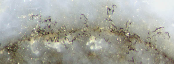
More glossy black matter of this type is shown in Figs.4,5.
 Figs.4,5: Black substance
near the contour of Aglaophyton
sections
on an old chert fracture face. Width of either picture 1.4mm.
Figs.4,5: Black substance
near the contour of Aglaophyton
sections
on an old chert fracture face. Width of either picture 1.4mm.
Obviously the black substance in Figs.1-5 had been
deposited after a horizontal crack had run along the chert layer. This
could have happened at any time within 400 million years, quite recent
times included. Unfortunately, the observations discussed here seem to
provide only little information suitable for narrowing this huge time
span.
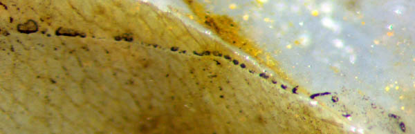
Fig.7. Flat blobs of black substance on a face
inclined to the picture plane. Width of the picture 2mm.
It
takes some effort to imagine the 3D-situation related to Fig.7. Let the
bluish area on the right be called the main fracture face of the
sample. The brown area on the left is the cylindrical replica of an
epidermis seen from within.
Here the crack had left the main
fracture face when it came upon the cuticle as an easy crack path.
The
string of black patches marks the intersection of the cylindrical face
with the imaginary continuation of the main
fracture face.
The patches are inside the chert behind the brown cylindrical face but
are out in the open on the bluish flat face. All of them are on the
common flat face, (which is not perfectly plane across the sample).
Fig.7 suggests that ...
- the main
fracture face once
had been one real crack with a narrow gap,
- black substance or its precurser
moved from the plant into the gap and arranged itself in peculiar ways,
- subsequently the crack more or less healed,
- later the chert layer broke into pieces, also
along the old "main fracture face",
- thereby the black substance was laid bare at the
surface, except for rare deviations as in Fig.7.
Apparently all
this could not have occurred quite recently but this does not mean that
it must have occurred in the distant past. (In this connection, one
should be careful not to confuse the black deposit of the rare glossy
type
considered here with the abundant black crusts, often circular ones,
recently formed on any stones, probably by cyanobacteria.)
Obviously the black substance or its precurser must have been in a
(transient) mobile or
fluid state. From the above observation (3) it may be concluded that
the
source of the possibly oily substance had been at the surface of the
embedded plants, which poses another problem
and suggests a solution. Usually the plants do
not
emit organic substances while or after being silicified. Something
unusual must have been going on. One thinkable
option is the following.
It
is known that the cuticle covering the epidermis is highly
decay-resistent so that it can persist in the chert over geological
timescales (and may make its presence felt as an easy crack
path as in Fig.7). It
consists of polymers and waxes which do not move about in the
chert. Perhaps the solid cuticle or some of its components can be
turned into mobile
or
fluid decay products by the action of microbes. In the present case,
the microbes could have reached the embedded plant through the gap of
the crack and broke down cuticle matter into fluid decay products
seeping out into the gap. Apparently a transient liquid substance is
seen in the
above pictures, with scattered dots of irregular
shape, which could be clusters of microbes. The comparatively big blobs
seem to have formed by accumulating some amount
of liquid of oil-like consistence, dots included, and by subsequent
transformation into a glossy black
solid substance.
It
must be admitted that this is not really an explanation since some
questions concerning the process steps have remained open.
If further investigations confirm the interpretation of these pictures
as evidence for the mobilisation of cuticle substance, they could serve
as illustrative material for the formation of mineral oil from plant
matter.
Photographs: Taken from the raw surface of a Rhynie
chert fragment found by Sieglinde
Weiss in 2014.
H.-J.
Weiss
2014
[1] H.-J.
Weiss :
Rhynie chert – Implications of new finds. European
Palaeobotany and Palynology Conference 2014, Padua.
 |
 |
70 |