Hollow
straws in pairs
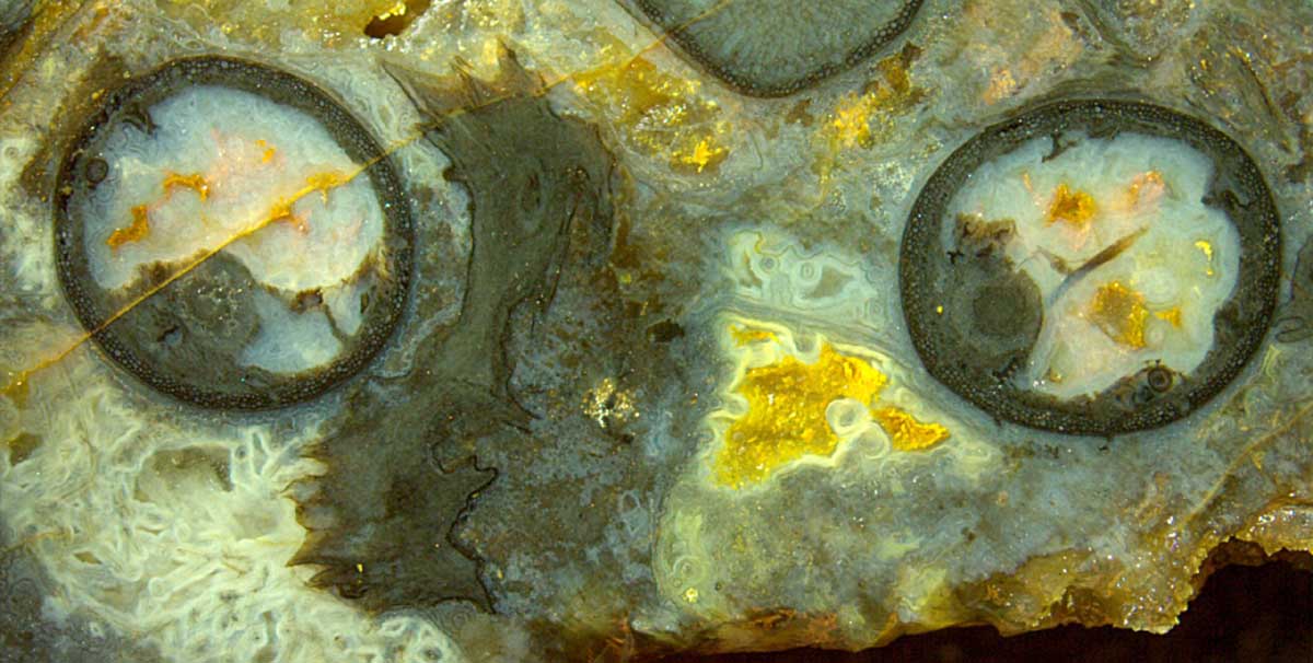
This picture of a cut and polished face of
a Rhynie chert sample offers more than sections of equal size and shape
near each other, nevertheless let us begin with this conspicuous
feature.
From
the rather similar aspect of the sections one can conclude that there
were two parallel shoots of equal size emerging
from a common base like prongs of a fork.
Their mode of degradation must have been
inherited from that base since the similarity could hardly have been
incidental.
The "hollow
straw aspect" seen on
either prong is often met with Aglaophyton.
Here it apparently supports the assumption
that this type of degradation had been predetermined or had formed in
the live plant, and
that
the plant was able to live with most of the cortex tissue missing,
provided
that a circumferential ring of tissue
and the central strand were still functioning.
As
it seems, this ring and the central strand were actively protected
against decay by unknown means. There
are still unsolved problems concerning these rings.
Image: Cross-section of forked Aglaophyton, either
prong with "hollow straw" aspect. Width 15mm.
The decay could have been brought about by some fungus spreading in the
cortex tissue of the live plant. Deformation and
beginning decay of cortex cells is seen
in the separate section above. Hyphae of an
aquatic fungus are
faintly visible as thin dark lines or dots surrounded by
whitish coatings, in the corner below left in this image. Two multiply
coated hypha cross-sections appear as "eyes " in a pale funny face near
the middle of this image.
The largely shrivelled and
shrunken plant section left of the middle indicates that there must
have been a quite different mode of decay, perhaps rotting of the
dead plant which had not become a
hollow straw.
Obviously the supply of dissolved silica had not been
sufficient to form silica gel and finally chalzedony throughout. There
were big and small pockets of water left. Some got distinct linings of
whitish or pale silica gel turning into chalzedony. Finally,
quartz crystals grew slowly in the remaining
cavities below right and at several
spots above.
Yellow or red stains of iron oxides had
been deposited in some of the small cavities above.
Apparently, iron salts and oxygen had not been there at the beginning,
otherwise a yellow or red chert would have formed.
Possibly the two components had entered into the water-filled cavities
and into the crack later by diffusion and combined there into
iron oxides.
Sample Rh2/230, Part 2
(39g), found by S. Weiss in 2014.
H.-J. Weiss
2018
 |
 |
130 |