Level fills in
hollow Aglaophyton
It is well known from small agates in volcanic rock that their
aspect can be quite different even though they may be only a few
millimeters apart, and that occasionally the banding or part of it is
horizontal. Similar formations, called here level fills for short, are
not rare in the Lower Devonan Rhynie
chert, which is silicified
water with plants, debris, and mud, where
former cavities due to gas bubbles or hollow plant parts became
gradually filled with silica clusters of various size which fused into
solid chalcedony later. Also it is known that the horizontal
bands sometimes thought
to be a relic of a water level in the cavity are no such.
The present small chert sample of 60g shows level
fills in Aglaophyton,
which is a not quite common sight: Fig.1. Obviously the fills vary
in colour and layer thickness. The same applies to the linings on
the cavity walls. This suggests that silica deposition
and gel formation had been highly sensitive to slight variations of the
concentration of substances, perhaps decay products, in the watery
silica solution.
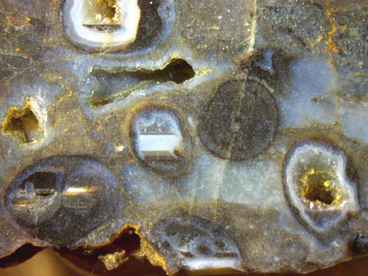
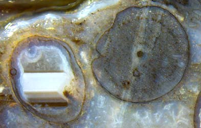
Fig.1: Rhynie chert with Aglaophyton
cross-sections, 5 hollow ones, now partially filled with a
series of silica deposits: linings and level layers of
chalzedony,
coarse quartz crystals as the final formation. Sample: Rh2/234, 60g, found by Sieglinde Weiss in 2014.
Width of the picture
18mm. See enlarged details below.
In the present sample, the level layers are there in the hollow straws
only but not in the other cavities. This suggests that the
watery suspension of tiny silica clusters, which settled down into a
"lake" with a distinct plane boundary against the clearer water above,
had formed preferably in the compartments where possibly some decay
products of the plant tissue were confined.
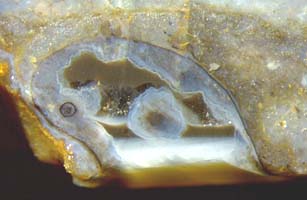
Figs.2-4: Aglaophyton sections,
largely hollow before silicification owing to decayed tissue.
(Fig.3
is 1cm outside the frame of Fig.1.) Height of the pictures 6mm.
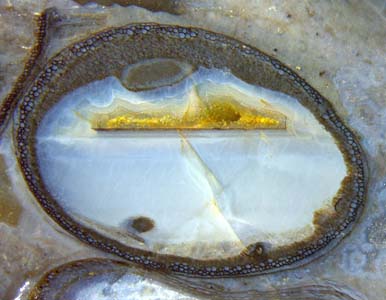
What appears as a dark fill in Fig.2 is only a thin dark plate of 40µm,
slightly inclined and thus seen from below through the clear quartz. A
similar but thinner plate, 20µm, is there in Fig3, also inclined but
not seen from below through the milky bluish chalcedony. Above the dark
plates, clear quartz crystals grew more slowly, with diameters up to
0.3mm, and part of the cavities has remained empty until now.
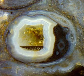
Fig.4 is more complex since there had been compartments where
silicification proceeded in different ways. What is seen as dark brown
on the
left is a compact fill with a plane light-coloured bottom in plain
sight because the space below is empty. The latter phenomenon is
unexpected but not very rare. What is now empty below a plane bottom of
a
deposit must have been a plane surface of another deposit which became
dissolved much later.
Much earlier when there were no deposits but only
water in the cavity of Fig.4, a fungus hypha had been there on the
left. When
the cavity got
its thick bluish lining, the hypha got its thick
bluish coating, which remained unaffected later while deposits
accumulated and vanished around it. It is still seen with a tiny dark
dot in the middle, which is the hypha cross-section. The bright bar on
the right is
a layer consisting
of
conspicuously glittering quartz crystals in the depth. Below it there
are smaller levels independent of those on the left.
Also the thin dark plate in Fig.2 must have been free-standing for a
long time after the deposit below had dissolved, similar as in Fig.4,
so that the space became
free for the slowly growing clear quartz which is there now.
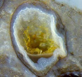 Figs.5-7:
Aglaophyton sections,
largely hollow before silicification owing to decayed tissue. Height of
the pictures 5mm, 4mm, 5mm.
Figs.5-7:
Aglaophyton sections,
largely hollow before silicification owing to decayed tissue. Height of
the pictures 5mm, 4mm, 5mm.
Same scale for Figs. 2-7.
The dark ring of cells indicating the presence of the symbiotic fungus
Glomites,
which is often found with Aglaophyton,
is
faintly seen on the well preserved cross-section of 4mm in Fig.5.
What
the fill in Fig.5 has in common with Figs.2,3 is the dark layer on top
of the stack,
here 90µm.
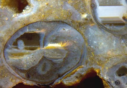
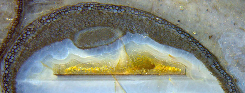 Fig.8
(below): Enlarged
part of Fig.3, hollow Aglaophyton not quite
filled by a series of silicification processes.
Fig.8
(below): Enlarged
part of Fig.3, hollow Aglaophyton not quite
filled by a series of silicification processes.
The enlarged
part of Fig.3 is to show that
the hollow plant parts with silica fill may offer rich detail
concerning
silicification and plant life as well. It suggests that later in its
life the
plant was able to do away with most of the cortex tissue and become a
live hollow straw, with a wall of well-preserved
cells
along the circumference, some degraded tissue inside, and the central
strand,
probably still functioning, leaning to one side.
The fills in Figs.6,7 are not plane on top. Hence, they had not been a
liquid suspension prior to solidification but
rather a slurry.
After the deposition of linings and layers from strongly supersaturated
solutions, quartz
crystals grew from slightly supersaturated
solutions in the cavities. Thereby the
cavities became completely filled in Figs.5,6 but remained
partially empty as in Fig.7.
So
it appears that much of what is seen in this small sample of Rhynie
chert with rich detail is compatible with the logic of silicification
but the cause of the
diversity remains enigmatic. Also the brown cloud in the upper part of
the former cavity in Fig.6, above the clear coarse quartz, does not
seem to fit in.
H.-J. Weiss
2015, 2018
 |
 |
75 |