Same symbiotic fungus in
Aglaophyton
and Rhynia
?
Aglaophyton
(former Rhynia major),
the most common plant in the Rhynie
chert, is often inflicted with an anomaly showing on cross-sections as
a concentric ring of cells with dark content scattered among the normal
cells, positioned at a depth of a few cell diameters below the
epidermis (Figs.1,2). High magnification reveals that the dark matter
in the cells consists of a dense tangle of tiny branched fungus hyphae,
much the same as in extant cases of symbiosis known as arbuscular
mycorrhiza [1]. The phenomenon has been described in detail and
explained as being due to the fungus Glomites rhyniensis
[1]. As a
remarkable fact, much thicker hyphae, and even thicker strands of
combined hyphae of this fungus, are often seen in the chert between the
plant matter even with low magnification. They must have grown in the
mud and water and developed the tiny variety of hyphae when entering
into the cells of the living plants.
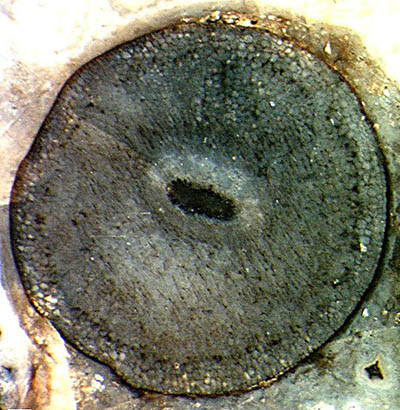
Figs.1,2: Aglaophyton
sections (4mm)
with dark cells affected by the fungus Glomites rhyniensis,
loosely
arranged in a layer below the surface.
According to [2], this fungus species seems to be restricted to
Aglaophyton (*).
So it is worth mentioning that cross-sections with the
same aspect, namely dark cells loosely arranged as a concentric ring,
are also seen in the similar but smaller plant in the Rhynie chert, Rhynia
gwynne-vaughanii (Fig.3 below, same scale as Figs.1,2).
This may not seem important in itself but taken together with other
recent observations it may serve as another argument in a scientific
dispute unfortunately started by a publication by David S. Edwards
[3]. 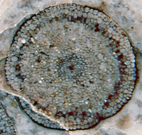 There
the plant then known as
Rhynia major [4] had been declared a
non-vascular plant, unrelated to the rhyniophytes but somehow related
to the mosses, and re-named Aglaophyton
major. This
name and
interpretation are presently officially valid although contrary
evidence has recently accumulated. (See, for example,
Rhynie
Chert News 2.)
The present observation, too,
seems to
indicate a closer
relationship between the two plants, as originally thought [4] and
apparently also assumed by Dianne
Edwards [8].
There
the plant then known as
Rhynia major [4] had been declared a
non-vascular plant, unrelated to the rhyniophytes but somehow related
to the mosses, and re-named Aglaophyton
major. This
name and
interpretation are presently officially valid although contrary
evidence has recently accumulated. (See, for example,
Rhynie
Chert News 2.)
The present observation, too,
seems to
indicate a closer
relationship between the two plants, as originally thought [4] and
apparently also assumed by Dianne
Edwards [8].
Fig.3 (right): Rhynia
section
(2mm) with dark cells whose aspect strongly resembles Aglaophyton
sections with Glomites
rhyniensis.
H.-J.
Weiss ;
2009, 2014
(*) Annotation: The
phenomenon of arbuscular mycorrhiza has
been mentioned in [5,6] for Rhynia,
too, without explicitly naming the
fungus. In [1,5,7] there is a rare
photograph of a tiny hypha
of Glomites rhyniensis
penetrating a cell wall of Aglaophyton.
(This picture is also seen here.)
[1] T.N.
Taylor et al.: Fossil arbuscular mycorrhizae from
the Early Devonian,
Mycologia 87(1995), 560-73.
[2] T.N.
Taylor et al.: Fungi from the Rhynie chert,
Trans. Roy. Soc. Edinburgh, Earth
Sciences 94(2004 for 2003), 457-73.
[3] David
S.
Edwards , Aglaophyton
major ..., Bot. J.
Linn. Soc. 93(1986), 173-204.
[4] R.
Kidston, W.H. Lang: On Old Red Sandstone
plants showing structure from the Rhynie Chert bed,
Part II, Trans. Roy. Soc. Edinburgh
52(1920), 603-27.
[5] H.Kerp:
De Onder-Devonische Rhynie Chert,
Grondboor & Hamer 58(2004),
33-51, Fig.19.
[6] T.N.
Taylor, E.L. Taylor: The Rhynie chert ecosystem: a model
for understanding fungal interactions,
in: Microbial Endophytes, eds.:
Ch.W.
Bacon, J.F. White Jr., Marcel Dekker Inc., New York 2000.
[7]
T.N. Taylor, E.L. Taylor, M. Krings:
Paleobotany,
Elsevier 2009, Fig.3.96.
[8] Dianne
Edwards : A review of the sporophytes of embryophytes
in the cherts at Rhynie,
Trans. Roy. Soc. Edinburgh, Earth
Sciences 94(2004 for 2003), 397-410.
 |
 |
32 |