Early land plant seedlings
As
a well known but peculiar fact, the "early land plants", more or less
well preserved in the famous Lower Devonian Rhynie chert, are seen
there abundantly as adult plants and as spores but very seldom as
germinating spores and occasionally as mm-size plants, as the one shown
in Rhynie
Chert News 25.
Possibly the early stages had been both short-lived and prone to fast
decay. Therefore, the rare chert samples with "baby plants" as seen in
Fig.1 deserve
particular attention. The plantlets are occasionally seen as a few
specimens scattered among large amounts of grown-up plants. So it has
come as a surprise that about a hundred mm-size plants have recently
been seen on the cut halves of a small sample of mere 60g. There they
seem to be restricted to a layer of 2cm height.
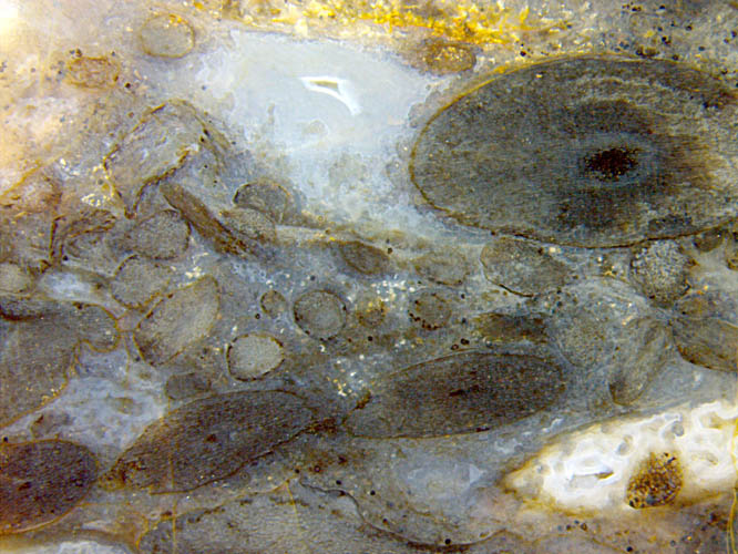
Fig.1:
Rhynie chert with plant sections: Aglaophyton
(above right), probably
Rhynia
(below), and randomly arranged mm-size seedlings of various
orientation. Width of the image 11mm.
As expected, they have got a cellular structure, which is indicated on
the plantlet enclosed in white chalzedony in
Fig.1, or seen
clearly with higher magnification (Figs.2-6). Apparently they do not
yet have
developed a central strand. Among the numerous randomly oriented
sections, those which incidentally are close to a plane
containing the central axis can most suitably give an impression of
the overall shape and a few details. As expected, the plantlets have
got a base and a top or front end. There is a texture in the tissue
near the base, with cells arranged in more or less diverging files
apparently originating from one point below, as in Fig.2. The
lowermost part seems to be broken off in Fig.3. Also in Fig.3, a layer
of cells on the left, set off from the tissue by a dark stain, looks
like an epidermis. The approximate axial symmetry, which is apparent
from nearly circular cross-sections as seen in Figs.1,5,6, may be
spoilt by
lateral bulges as in Figs.2,4. It has not been found out whether such
bulges mark the onset of lateral branching, as suspected in a previous
contribution, or the places where rhizoids grow from, as suggested by
Fig.4.
Fig.2 (below left): Plantlet, 1.4mm, with upward diverging cell files
at the bottom and two lateral bulges.
Fig.3 (next): Plantlet, 1.35mm, with stained epidermis and upward
diverging cell files at the bottom.
Fig.4: Plantlet, 2mm, with upward directed cell
files in the middle, rhizoids on a lateral
bulge and below.
Note also
a few spores (70µm) opening along their trilete mark.
All
figures show the same sample. Figs.2-6
are of the same scale.
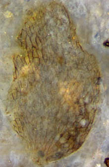
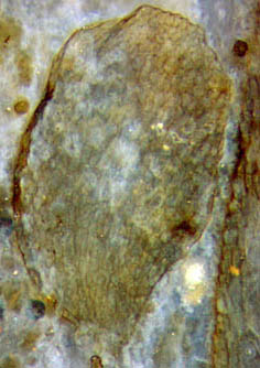
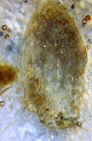
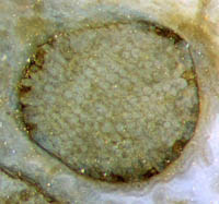
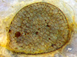
Fig.5 (above): Plantlet cross-section, width 0.7mm. Note
the homogeneous tissue structure.
Fig.6 (next): Plantlet
cross-section, width 0.9mm, with clearly seen epidermis above.
It
is hoped that finds of the kind described here can eventually
contribute to progress on the way to a solution of the intriguing
gametophyte /sporophyte problem of the early land plants in the Rhynie
chert. The gametophytes seem to have been seldom preserved. Images [1]
of much younger ones (up to 0.1mm) and much older ones (cm-size) are
available for comparison with own finds but of no much use here. (Those
images have been reproduced in [2] as "Courtesy H. Kerp", some with
erroneous size data.)
Archegonia
are known to occasionally appear as dark spots sunken into the surface,
superficially resembling the spots in Figs.1,5, but closer inspection
reveals that the latter probably are cells affected by some
fungus or microbe
as it is also seen on adult Aglaophyton
in this sample.
There is one feature which seems to be in favour of a
sporophyte interpretation: The lower end of the elongate plantlets
often looks as if broken off somewhere (Figs.2,3). They might
have grown from fertilized egg cells still sitting on the
gametophyte, then become detached while the gametophyte decayed fast.
It would be interesting to know
at which stage of growth a central strand is formed. Perhaps the
faintly seen files of elongate cells in Fig.4 and a cluster of
small cells near the middle in Fig.6 are first indications of such
process.
H.-J.
Weiss
2015
[1] W. Remy, H. Hass: New information on gametophytes and sporophytes of Aglaophyton ... Rev. Paleobot. Palyn. 90 (1996), 175-193.
[2]
T.N. Taylor,
E.L. Taylor, M. Krings: Paleobotany,
Elsevier 2009, 232-244.
 |
 |
88 |