Conspicuous in the chert - synangia of
Scolecopteris
Parts of the large fronds of the Psaronius
* tree ferns
are well-known fossils
from several locations worldwide, and they are abundant in some of the
chert variants from the Lower Permian Döhlen basin. When preserved
3-dimensionally,
as in chert, they are called Scolecopteris,
else Pecopteris.
As a
characteristic feature they bear groups of sporangia fused at their
base, called synangia, on the lower side of the pinnules. The build of
the synangia is not uniform among the species of Scolecopteris:
They may be thin-walled and largely
enveloped by the recurved margin of the pinnule and thus protected
(Fig.1), or
free-standing and thick-walled (Fig.2), thus protecting themselves, or
something
in between. The more
thick-walled forms were probably hard and dry like the seed capsules of
many extant flowering plants and therefore decaying more slowly than
the pinnules bearing them. Therefore the synangia
are often the most distinctly
seen parts of the whole plant. This should make them suitable for
the identification of species.
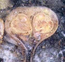
Fig.1: Scolecopteris
pinnule cross-section with synangia protected by fringes extending from
the pinnule margins, seen on the natural surface of a chert
layer fragment found near
Perdasdefogu, Sardinia, and resembling Sc. elegans, a
common species from Döhlen basin. Pinnule width
1.7mm.
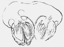
Fig.2: Scolecopteris
pinnule cross-section with exposed synangia composed of
thick-walled sporangia, less-common variety from
Döhlen basin, drawing after polished chert face. Width of the pinnule
1.6mm.
The identification of species by their
synangia turns out less practicable than
expected, with the
problems being partly genuine and partly generated by palaeobotany
itself. The scarce fossil material available to some researchers in
this
field led to the statement that the synangia of the "maggot fern"
Scolecopteris elegans
were composed of 4 to 5 sporangia and had "radial" or "bilateral"
symmetry. Such
statements, by repeated quotation, have found their way into the latest
publications
on the subject [1,2,3] despite of glaring evidence for the contrary:
Symmetry is no useful
concept here since synangia of various symmetry types, including no
symmetry at all, are usually found together on one pinnule (Figs.3,4 ),
and the
number of sporangia varies between 3 and 6 (Figs.3-8), with rare cases
of 2 and 7.
This should have been a well-known fact since the early work by Zenker
[4], who found the absence of a common symmetry so remarkable that he
drew a variety of synangium cross-sections, including non-symmetrical
ones.
Zenker
was readily referred to for the introduction of the name Scolecopteris
but apparently his paper [4] was not thoroughly inspected.
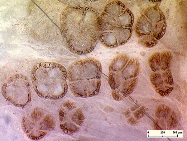
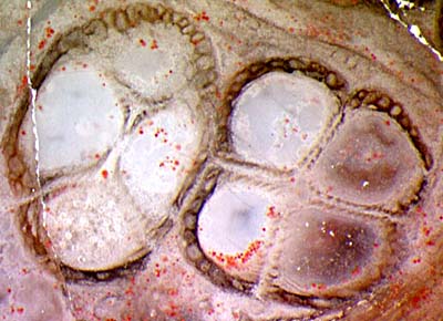
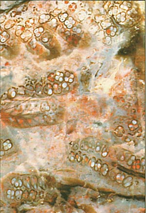
Fig.3,4: Usual aspect of Scolecopteris
synangia in cross-section: symmetry of various
type or absent. Photographs: H. Sahm.
Fig.3 (left): The two rows of synangia below are on one
pinnule. They
have been cut more or less near their tips.
Fig.4: With of the picture 1.23mm.
Another inappropriate notion surviving in the literature on Scolecopteris is
that of "spindle-shaped" or fusiform sporangia [3] although the
cross-sections of
the sporangia are not even approximately circular, as seen here.
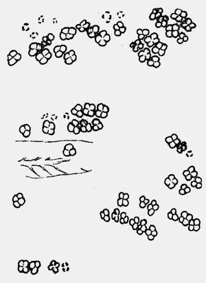
 Figs.5,6: Chert face with
conspicuous array of Scolecopteris
synangia cross-sections on pinnules hardly visible
owing to decay [5]. Only the well seen cross-sections in Fig.5 have
been included into the drawing. Width of the picture 9mm. Photograph:
W. Schwarz.
Figs.5,6: Chert face with
conspicuous array of Scolecopteris
synangia cross-sections on pinnules hardly visible
owing to decay [5]. Only the well seen cross-sections in Fig.5 have
been included into the drawing. Width of the picture 9mm. Photograph:
W. Schwarz.
The good visibility of the synangia in Fig.5 may be due to
the higher decay resistence of the possibly dry and hard sporangium
walls compared to the soft pinnule tissue. There is something else
which is remarkable about Fig.5: Among the 65 synangia
seen here in cross-section, 25 are less-common ones with only 3
sporangia,
and none is seen with 5.
The large number of synangia can easily mislead to the guess that Fig.5
is possibly a
representative part of the sample with a particular
species
whose synangia are composed of 3 and 4 sporangia only. However, there
are
synangia with 5 sporangia outside the frame of the picture, which may
serve as a warning not to draw conclusions hastily. This applies also
to the preliminary statistics in the table below, based on 69 chert
samples with synangia seen in cross-section.
The
numbers n of the n-fold synangia seen in one chert sample are listed in
the first line of the table. The second line is the number of related
samples.
3 4 3 4
5 3 4 5 6
4 4
5 4 5
6 4 5 6 7 5
6 5 6
7
14
10 3
15
19 5
1
1
1
Note that this does not mean that a type characterized by 4-fold
synangia only has been
found in 15 samples. Additional cut faces would
probably provide synangia
with n = 3 or 5 .
As
a general observation from the Döhlen basin, synangia with 4, 5, and 6
sporangia are often seen in close vicinity, on the same or neighbouring
pinnule (Fig.7).
Most
sporangia were empty when the fronds or pinnae fell into the water and
became silicified but some were still filled with spores (Fig.8). Here,
the spores are not lying in the sporangia loosely but are seen to be
still arranged in some original order. As another remarkable feature of
the sporangia in Fig.8, the walls have
completely collapsed where they mutually touch so
that they are seen in cross-section as a mere thin line. (Compare
Figs.4,7 where the individual walls are still seen.)
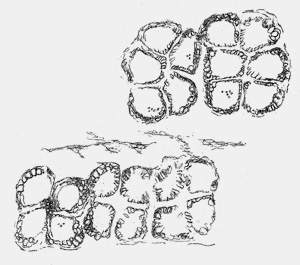
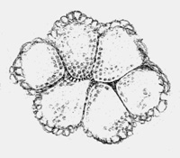
Fig.7 (far left): Scolecopteris
synangia of different type on neighbouring pinnules, as seen on the raw
surface of a chert sample. Width of the picture
2.2mm.
Fig.8: Synangium with sporangia completely filled with immature spores
and with walls partially collapsed into thin foils. Width
of the synangium 0.9mm.
The
observations lead to the conclusion that notions like symmetry of the
synangia and spindle shape of the sporangia are not helpful for the
identification of Scolecopteris
elegans
and for differentiation between similar strains closely related to it.
"Rotational symmetry", which means an n-fold rotational axis in the
synangium, is a characteristic feature of a group of Scolecopteris species
including Sc. globiforma
and Sc. unita
[6] but not of Sc.
elegans.
Other features of
the synangia are more suitable for differentiation between maggot fern
variants or species:
The pedicel
is usually much shorter than wide and thus hardly visible, or
it can be
rather conspicuous. The sporangia can be thick and short or narrow and
tapering. Hairs on the
sporangia are well known from Scolecopteris
species found in "coal
balls" in North America [7] but have been found in the Döhlen basin in
only two samples hitherto. An evaluation of
the more useful but rare or elusive features of the synangia in the
cherts of the Döhlen basin has not yet been done. Considering that lots
of chert samples with Scolecopteris recovered
in the 1990s have been stored away and not even looked at, it is well
possible that some correlation between synangia features and fern
variants will be found when the samples are
thoroughly investigated.
Samples: own finds if not indicated
otherwise.
Figs. 2-8:
Döhlen
basin, Freital
Fig.1: Pd/2.1, Perdasdefogu, Sardinia, 2009,
Fig.2: Bu4/31.1, found in 1996 on
the property of Lippert,
Burgk,
Bernhardts Weg 25.
Fig.3-6: Bu10/7, provided by H.
Nitzsche, Burgk,
Kohlenstr. 24, in
1998.
Fig.7: Bu8/18,
found in 1997 at Burgk,
Am Seilerschuppen,
and kept by U.
Wagner.
Fig.8: Bu2/5.1, found
in 1995 as one of the
first own finds on the "maggot stone" site,
on the property of W. Netzschwitz Kleinnaundorf,
Kohlenstr. 23, and
kept there.
* Uncommonly big and well-preserved specimens
of Psaronius,
the stem of several "maggot fern" species, are on display
at the Naturkunde Museum Chemnitz.
H.-J.
Weiss 2011, amended 2012
[1] M. Barthel,
W. Reichel, H.-J. Weiss: "Madensteine" in Sachsen.
Abhandl. Staatl. Mus. Mineral. Geol. Dresden 41(1995),
117-135.
[2] R.
Rößler : Der versteinerte Wald von Chemnitz, 2001.
[3] M. Barthel:
The maggot
stones from Windberg ridge, Germany.
in:
U. Dernbach, W.D.
Tidwell: Secrets
of Petrified Plants. D'ORO Publ. 2002. p65-77.
[4] E.
Zenker:
Scolecopteris elegans,
ein neues fossiles Farrngewächs mit
Fructification. Linnaea 11(1837), 509-12.
[5] H.-J. Weiss:
Beobachtungen zur Variabilität der Synangien des Madenfarns. Veröff.
Museum f. Naturkunde Chemnitz 25(2002), 57-62.
[6] M.A.
Millay, J. Galtier: Studies
of paleozoic marattialean ferns: Scolecopteris globiforma from the
Stephanian of France.
Rev. Palaeobot. Palyn. 63(1990), 163-171.
[7] M.A. Millay:
Study of paleozoic marattialeans. A monograph of the American species
of Scolecopteris.
Palaeontographica B169(1979),
1-69.
|
 |
 5 5 |