Trigonotarbids: known for a long while but still
surprising
Trigonotarbids are extinct little predatory creatures related to the
spiders, mites, and scorpions. They resemble spiders in habit and
behaviour but did not produce silk. (For details and reconstruction see
[1].) Their exuviae shed in moulting are well-known fossils in the
Rhynie
chert. They are incidentally seen on cut faces (Fig.1) and in
rare cases as a relief on the surface of the chert sample (Fig.2).
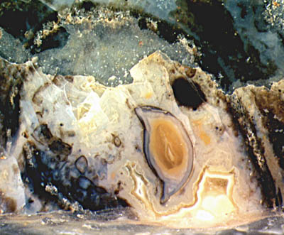
Fig.1: Exuviae silicified within a
hollow straw of
Aglaophyton
where the trigonotarbid probably hid for moulting; body
cross section with agate banding, width 2mm;
disarranged legs and other parts nearby. Width of the picture 6mm.
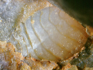
Fig.2: Trigonotarbid body, ventral
side, with distinctly seen superficial
segmentation, on the raw outside of a Rhynie chert sample. Width
of the picture 3.4mm.
Fig.3 (below): Moult of trigonotarbid leg
consisting of 7 parts, with bristles. Width of the picture 1.5 mm.
Photographs
(Figs.1,3) by H.
Sahm .
As a
primitive feature, the bulky body shows the superficial segmentation
which has been lost in nearly all extant spiders. The articulated hairy
legs are made up of the same parts as the spiders' (Fig.3).
In addition to the three species identified in the chert hitherto there
are rare sightings of comparatively huge specimens, as a still
undescribed one shown in [2]. One of those which are much larger than
usual is seen in lengthwise section in Fig.4. As it is
preserved as a whole among prostrate shoots and an immature sporangium
of Aglaophyton,
it was probably drowned in the flooding which also
flattened
the vegetation.
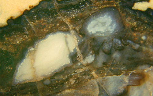
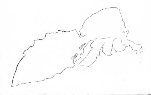
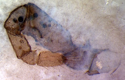
Fig.4: Lengthwise section and related drawing of an
uncommonly large trigonotarbid
among flattened vegetation, total length without legs 9mm. Note the
very faintly seen level bands inside the body
parts near the bottom,
suggesting preservation in a natural position. Width of the picture
10mm.
Fig.5 (below far left): Book lung with lamellae attached
to the enclosure on the left; detail of Fig.4. Width of the
picture 0.35mm.
Photograph by Th.
Hanke.
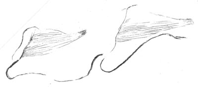
Fig.6 (below): Book lungs in
each of the first two segments of the rear part of
the body, see drawing of Fig.4;. Width
of the picture 1.7mm.
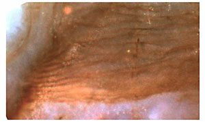
 Surprisingly,
the so-called book lungs made up of a stack of lamellae
are well preserved (Fig.5). Pictures of trigonotarbid book lungs have
been shown before [1,2] but seem to be scarce, and the present one
reveals some detail not seen in the aforementioned ones: There are two
stacks of lamellae, in fact two lungs, one behind the other (Fig.6), as
a relic of the more distinct segmentation of the trigonotarbids'
ancestors.
Surprisingly,
the so-called book lungs made up of a stack of lamellae
are well preserved (Fig.5). Pictures of trigonotarbid book lungs have
been shown before [1,2] but seem to be scarce, and the present one
reveals some detail not seen in the aforementioned ones: There are two
stacks of lamellae, in fact two lungs, one behind the other (Fig.6), as
a relic of the more distinct segmentation of the trigonotarbids'
ancestors.
Remarkable is also the apparently narrow waist as seen on the outline
in Fig.4 since the body of trigonotarbids allegedly does not
look clearly subdivided into a front and rear part.
Annotation 2015: The specimen in Fig.4 possibly
represents a less advanced trigonotarbid species
with a waist as an ancient feature. (See also Rhynie Chert
News 42
.)
The pictures have been taken from 4 Rhynie chert samples.
H.-J. Weiss
2005 2015
[1] www.abdn.ac.uk/rhynie
[2] H.
Kerp, H. Hass : De Onder-Devonische Rhynie Chert,
Grondboor & Hamer 58(2004),
33-51.
 |
 |
9 |