Trichopherophyton
and Ventarura
in one chert sample
The mere fact that the two rarest and therefore last
discovered plants [1,2] in the Lower Devonian Rhynie chert have been
found
together in one piece of chert seems worth mentioning.
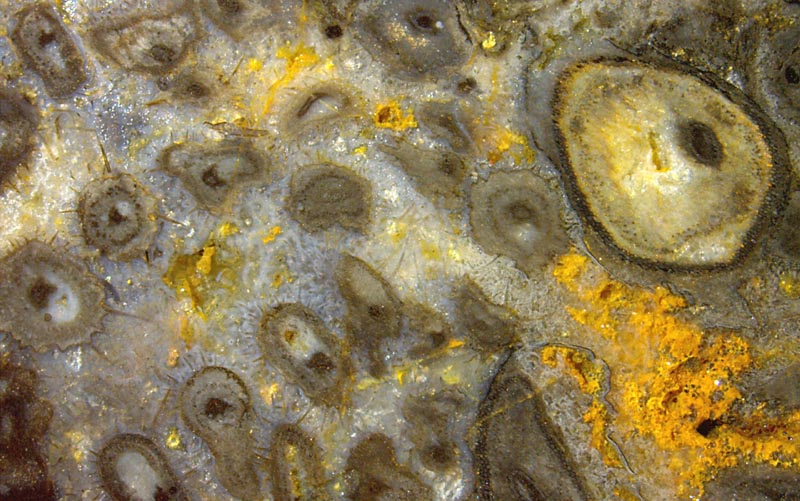
Fig.1
(right): Cross-sections of more or less deformed shoots of Ventarura
and smaller Trichopherophyton
with bristles. Width of the image 17mm.
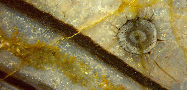
Fig.2 (below): Cross-section of well-preserved Trichopherophyton
beside lengthwise section of Ventarura.
Width of the image 8mm.
Trichopherophyton
is easily
recognized here by its bristles
but often it is seen without. (See Rhynie
Chert News 49,
Fig.3.) Then it may be distinguished from Rhynia,
whose diameter is in the same range, by the scalariform pattern on the
tracheid walls seen on longitudinal cuts of the xylem strand.
Apparently the bristles
served as a protection
of the sporangia against spore-eating crawling creatures. This
is
suggested by the fact that the bristles are borne only
on the
upper parts of the plant. No other species in this Lower
Devonian habitat had developed such
protection. Unlike other plants in the chert, Trichopherophyton
is often seen in
good shape
and with well-preserved tissue, as in Fig.2.
Ventarura
is the bigger one, as seen in
Fig.1 above right and in Fig.2 below left. Its upper parts are
distinguished by a unique feature of a quite different kind: It is a
big tube of rot-resistant tissue appearing on cross-sections as a ring
with clearly visible cell structure, most often with dark aspect, which
gives the impression of strength and has misled some observers to an
interpretation as sclerenchyma. Such interpretation has been rejected
by the observation that the cell walls are normally thin but appear
thick owing to a dark coating,
probably of microbial origin and grown while the dead plant was
decaying in the swamp water. (See Rhynie
Chert News 60,
Fig.6.) Occasionally the tube is the only part
of Ventarura that
is seen, as in the case of the lengthwise section in Fig.2. Apart
from its conspicuous appearance in the chert, the persistent tube must
have had a purpose in the live plant. A purpose, however, is not
obvious. With its position inside the cortex tissue it would not have
protected against damage to the surface. As a far-fetched
interpretation, the tube, if poisonous or tasting bad, could have
prevented sap-sucking creatures from gnawing towards the phloem
surrounding
the xylem strand.
The two rare plants were first seen together in one sample in 2014.
H.-J. Weiss
2016
[1]
A.G. Lyon, D. Edwards: The first Zosterophyll from the
Lower
Devonian Rhynie Chert.
Trans. Roy. Soc. Edinburgh, Earth Sci.
82(1991), 323.
[2]
C.L. Powell, D. Edwards, N.H. Trewin: A new vascular
plant from the Lower Devonian Windyfield chert, Rhynie,
Trans. Roy. Soc.
Edingurgh, Earth Sci. 90(2000 for 1999), 331-349.
 |
 |
95 |