Nematoplexus
with a wry twist ?
When trying to find out something new about a fossil, one need not
always
have the very specimen at hand. Sometimes it is sufficient to have a
published picture and look for features not noticed by the authors.
(See Rhynie
Chert News
8, 28, 29.) The picture in Fig.1 can
serve as another example.
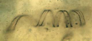
Fig.1: Peculiar object described in [4] as "a spirally coiled
smooth-walled tube of Nematoplexus".
Width of the picture 235µm. Copyright owned by University
Münster.
Coil diameter 40µm.
A few introductory explanations are appropriate here. Nematoplexus [1]
is one of a few Silurian – Lower Devonian fossils denoted nematophytes
and lately listed among "Enigmatic Organisms" [2]. It is
made up of a tangle of wound tubes probably embedded in gel. It is a
rare fossil, apparently known from only 4 samples of Rhynie chert,
and only a few pictures are available. The type specimen found by Lyon
[1] had been photographed anew with advanced equipment [3]. Two of the
pictures are shown on the website of Aberdeen University [4].
Another specimen of Nematoplexus
is pictured in [5], where it is not
specified as such but presented merely as a nematophyte. It was dug
from a separate stratum near Rhynie whose chert pods, according to [5],
are named
Windyfield chert.
The tubes of the own specimen, seen in Fig. 2, have been inspected and
photographed on the raw surface of the sample. They look like those in
[1] and [5] except for the size. (Note that this picture should have
been reproduced twice as large to obtain the same magnification as
Fig.1. See Rhynie
Chert News
29.)
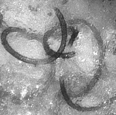
Fig.2: Three Nematoplexus
spirals with right-hand thread, cut off at the sample surface,
own sample with bigger variety of Nematoplexus
(new species ?), coil
diameter 190µm.
The spiral below right comes out of the surface near the centre of
the picture so that part of it is cut away and hence not seen, which
may
be confusing when one tries to see the 3D-structure. Width of the
picture 350µm. Photograph by C.
Kamenz,
2009.
It must be mentioned that the thread of virtually all Nematoplexus
filaments pictured so far is right-handed, and therefore it is felt
that any exception to this rule might hint at something interesting.
Even with the very few pictures available for comparison, it can be
stated that the wound filament in Fig.1 is out of the ordinary in more
than one way. First, it rather looks like a ribbon than like a tube.
Well, a collapsed tube could become a ribbon but this one seems to be
the only one which underwent such collaps. Second, the diameter of the
thread or coil in Fig.1 is as small as 40µm while it is usually
80...120µm or up to 190µm in Fig. 2. Third, the type of thread of the
wound filament seems to change suddenly from left-handed to
right-handed, as if a screw and its mirror image had been joined. This
peculiarity in Fig.1 was not noticed before as none of the authors
[1-5] considered the type of thread.
The rare deviation from right-handedness gives rise to the question of
how it could be brought about. Imagine, for simplification, a ring with
a
cut: By displacing the ends out of plane this or that way, a
left-handed or right-handed thread is formed, or rather one turn of a
thread or coil. Likewise, a short thread fragment could change its
screw type
by some incidental deformation, which would explain the presence of an
occasional left-handed short fragment, or end region of a longer
filament, among right-handed ones. However, such explanation does not
apply to the peculiar case seen in Fig.1. There remains the suspicion
that the apparently obvious change of the screw type is an optical
illusion due to the limited depth of focus. Here, only another
inspection of the sample could provide an answer.
As another way out, the object in Fig.1 might be no collapsed tube but
a ribbon, perhaps a spiral wall thickening left over from a decayed
tube of about 50µm diameter, a size which would not be compatible with
Nematoplexus
as we know it but is not uncommon among nematophytes. (See
Rhynie
Chert News
30.)
It is the aim of this essay to point out that there is a problem
with Nematoplexus
which was not noticed before and which does not seem
to have a simple solution. As with any tough problem, one can rightly
expect something interesting behind it, hence it deserves attention.
With some preparational effort, the samples presently available could
provide more pictures of wound filaments, which might help to solve
the problem posed by Fig.1. Further it can be expected that more
specimens of the elusive Nematoplexus
will be discovered, also by
thorough inspection of the Rhynie chert samples already stored in
collections.
H.-J. Weiss
2010
[1]
A.G. Lyon:
On the
fragmentary remains of an organism referable to the nematophytales from
the Rhynie chert,
Nematoplexus rhyniensis,
Trans.
Roy. Soc. Edinburgh 65(1961-62), 79-87, 2 tables.
[2]
T.N. Taylor,
E.L.Taylor, M. Krings: Paleobotany, Elsevier 2009.
[3] Palaeobotanical Research Group, University Münster.
[4] www.abdn.ac.uk/rhynie/nemato.htm
[5] S.R.
Fayers, N.H. Trewin: A review of
the palaeoinvironments and biota of the Windyfield chert,
Trans. Roy. Soc. Edinburgh, Earth
Sci., 94(2004 for 2003), 325-339.
 |
 |
39 |