A Lower Devonian alga resembling the
xanthophyte Botrydium
 The
bunch of funnel-shaped organisms in a small sample of Rhynie chert
(Fig.1) has come as a surprise as it differs from any known fossil from
that site. Apparently it is neither land plant, green alga, fungus, nor
microbial formation.
It resembles a xanthophyte, and if it really is
one, it would be, according to [1] and other sources, the
first fossil xanthophyte ever seen, since "There are no positively
known fossils of the Xanthophyta, ...". To explain the
reason why, [1] is quoted here again: "It is likely that the lack
of a fossil record results more from the fact that their cyst
morphology is
poorly studied, rather than from a lack of preservation, since the
silicified cysts of this group ought to be found."
The
bunch of funnel-shaped organisms in a small sample of Rhynie chert
(Fig.1) has come as a surprise as it differs from any known fossil from
that site. Apparently it is neither land plant, green alga, fungus, nor
microbial formation.
It resembles a xanthophyte, and if it really is
one, it would be, according to [1] and other sources, the
first fossil xanthophyte ever seen, since "There are no positively
known fossils of the Xanthophyta, ...". To explain the
reason why, [1] is quoted here again: "It is likely that the lack
of a fossil record results more from the fact that their cyst
morphology is
poorly studied, rather than from a lack of preservation, since the
silicified cysts of this group ought to be found."
This plain situation, without any fossil xanthophytes
through the ages, has apparently ended
with the discovery of Palaeovaucheria
[3], an alga
more than twice as old as the Rhynie chert, interpreted as a
xanthophyte in a "Gongrosira
phase".
Disregarding that dubious fossil from the Proterozoic, one can assume
that the silicified cysts which ought to be found according to
[1] have been found now.
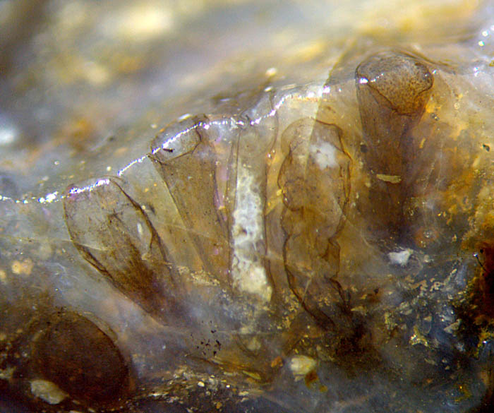
There are 8 funnel-shaped bags in this sample,
5 of which are seen here, more or less inclined towards the picture
plane. Because of this inclination, their bases
are deeper inside the sample and
therefore not seen.
Fig.1: Bunch of funnel-shaped bags seen at the edge of a Rhynie chert
fragment, resembling truncated cysts of the extant xanthophyte Botrydium. Width of
the picture 5mm.
From the outlines seen on the fracture faces it
may be concluded that the bags or cysts had nearly axial symmetry. One
of the bags,
2nd from the right, is the only one which had been subjected to
slight vertical compression, the result of which indicates that its
wall had
been very flexible and thin, estimated 1µm or less. The
top of the other cysts is missing, broken off when the small fragment
was shaped by disintegration of the chert layer.
Two not quite plane fracture faces meet at
an angle of about 100° in Fig.1, hence their common (slightly rounded)
edge is not quite straight, seen as a bright line
due to reflection of incident light. The picture plane has been chosen
such that it
essentially coincides with that fracture face which reveals most of the
bags, either as sections or as views through the transparent chalzedony.
Incidentally the two fracture faces are in such positions that they
permit
inspection of the bag on the right from different directions. Since
this bag is slightly deformed, the contour on one sample face is
irregular (Fig.2)
but not smoothly curved as it would be expected from a plane cut of an
inverted cone. Above the rounded edge in Fig.2,
which is marked by the bright reflections, the other face offers
a top view with partially circular outline (Fig.3),
except for the broken-off part. (To obtain a 3D-imagination, compare
Fig.2 and Fig.3, with Fig.1.)
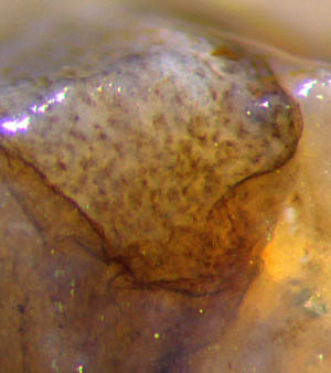
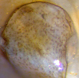
Fig.2 (far left): Detail on the sample front face cutting
sideways into
the funnel-shaped bag on the right in Fig.1, irregular outline due to
local deformation before silicification. Width of the picture 0.8mm.
Fig.3: Top view of the truncated cyst seen sideways in Fig.2. Width
of the picture 0.73mm.
The above claim of the discovery of a fossil
xanthophyte is substantiated by details from these images. An epidermis
is conspicuously absent, and so is any tissue, although the
interior of some bags is clearly seen to be filled rather evenly with
fuzzy dots of about 20µm. Other bags are empty. This is compatible with
Botrydium,
which is characterized by large numbers of nuclei and
chromatophores within a giant cell attached to moist soil [2]. When the
place is flooded, the nuclei and plasma turn into zoospores which
escape through an opening. Judging from the layering of the Rhynie
chert, flooding had occured repeatedly in the habitat which brought
forth the chert, which may
explain why some of the bags in Fig.1 are empty.
 The apparently
shapeless dots in Figs.2-4 had possibly been in some stages of
transformation into
zoospores when being silicified. As another option, the giant
cells of Botrydium
can become empty by producing and releasing gametes [2].
The apparently
shapeless dots in Figs.2-4 had possibly been in some stages of
transformation into
zoospores when being silicified. As another option, the giant
cells of Botrydium
can become empty by producing and releasing gametes [2].
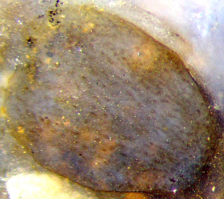
Fig.4 (right): One of the bags in Rhynie chert resembling cysts
of the extant xanthophyte Botrydium,
inclined section on the fracture face in
Fig.1 below left, seen here filled with nuclei (and
chromatophores ?).
Width of the bag 0.75mm.
Preservation in Rhynie chert had been governed by the competition
of decay and silicification. In the present case, another process had
also been relevant: Flooding could have initiated the formation and
release of zoospores or
gametes, similar as with extant Botrydium
[2]. Silicification interfered with this process so that the motile
unicells managed to escape from some of the tubes in Fig.1 before
silica gel formation but not from others. Hence, one should anticipate
the presence of fossil bunches of empty cysts which could easily be
misinterpreted as land plant cuticles.
Finally it can be assumed that, disregarding the
Proterozoic, the above images show the
first
fossil
xanthophyte, representing a 400-million-years-old one out of
this class of algae with about 600 extant species [1].
Photographs taken from the raw surface of a Rhynie
chert fragment of 20g found by Sieglinde
Weiss in 2014, stored under the
label Rh2/200.
H.-J.
Weiss
2014, modified
2016, 2020
[1] Introduction to the Xanthophyta,
www.ucmp.berkeley.edu/chromista/xanthophyta.html
[2] R. E. Lee: Phycology, p417. Cambridge 2008.
[3] N.J. Butterfield: A vaucheriacean alga
from the middle Neoproterozoic of Spitsbergen.
Paleobotany 30(2004), 231-252.
 |
 |
69 |