Nematoplexus
- Where the spirals come from
 The enigmatic "branch-knots", scattered among the
spiralling tubes
of the enigmatic organism
Nematoplexus seldom found in the famous Rhynie chert [1],
have
been thought to be places where the tubes branch profusely although a
spiralling tube has never been seen
branching.
The enigmatic "branch-knots", scattered among the
spiralling tubes
of the enigmatic organism
Nematoplexus seldom found in the famous Rhynie chert [1],
have
been thought to be places where the tubes branch profusely although a
spiralling tube has never been seen
branching.
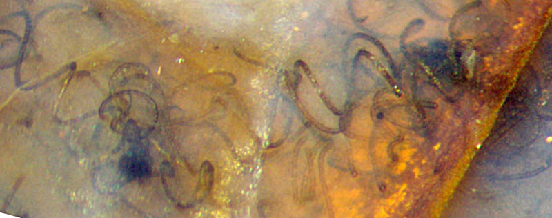
Fig.1: Spiralling tubes of Nematoplexus
and related "knots".
Image
width 1.3mm.
As seen in Fig.1 (reproduced from Rhynie
Chert News 71
),
the tangle of randomly distributed
tubes with dark "knots" in between is confusing, all the more so since
straight or weakly curved tubes may also be present: Rhynie
Chert News 51,
71 .
Higher magnification and
other
illumination have revealed that the knot on the right seems to consist
of two neighbouring ones. A
small protruding
part of the big black
knot in the depth of Fig.1 on the right is seen on the left of Fig.2.
(Note
that Fig.2 is turned to the left with respect to Fig.1.)
By lucky coincidence,
a small separate knot, not noticed in Fig.1 but seen in the middle of
Fig.2 now, is divided by a tilted crack plane
reflecting the incident light so that half
the knot, left of the divide, is illuminated from below while the other
half, right of the divide, keeps
in the shade. (The
off-white angular mineral platelet can serve as a mark for
comparison with Fig.1. The
bright white spot below is an irrelevant light reflex
from applied oil.) The colourful illumination is due to a
thin deposit of iron oxide on the reflecting crack face. 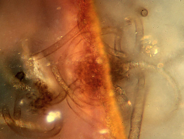
Fig.2: Small "knot" incidentally divided by a crack in the
chert reflecting the incident light. Image
width 0.3mm. Photograph by Gerd
Schmahl.
Most interesting is the junction of an
11µm-tube to what seems to be the indefinite
surface of the knot: Contrary to the common belief due to
the established term "branch-knot",
the tube is not produced by branching. The same
can be expected from the other tubes. One tube right of the divide, for
example, is
wider than normal where it emerges from the knot.
In addition to the tube diameters of 10-12.5µm
in Fig.2, there is only one clearly seen 4µm-tube
and a straight one with
17µm.
(More non-spiralling tubes are present at other places in this sample.)
Finally it can be stated that the
spiralling tubes of
Nematoplexus seem
to emerge
from the so-called branch-knots
without any branching.
Sample: Rh15/79, Part 4, obtained
from Barron in
2014.
Annotation
2020: For a
discusion of the subject in a wider context see Rhynie
Chert News 152.
H.-J. Weiss
2018 2020
[1] A.G.
Lyon: On the fragmentary remains of an organism referable
to the nematophytales, Nematoplexus rhyniensis.
Trans. Roy. Soc. Edinburgh
65(1961-62), 79-87, 2 plates.
(Scale error on Plate I Fig.1: not x19
but x1.8)
 |
 |
134 |