Ventarura
- peculiar and confusing
Every one of the seven early land plants hitherto discovered in the
Lower Devonian chert from
Rhynie, Scotland, is characterized by at least one unique feature.
Plant cross-sections with a dark concentric ring at some distance from
the periphery and consisting of rot-resistant cell walls are
characteristic of the lately discovered Ventarura
[1].
The ring seen on cross-sections is, of course, a tube inside the stem.
Unfortunately, reality is not as simple as this: The characteristic
feature may be a ring segment instead of a closed ring, it may be pale
instead of dark, and it may be so close to the periphery that the whole
can be mistaken for a hollow straw of the most abundant
plant in the
Rhynie chert, Aglaophyton.
Resulting from a peculiar growth mode, the originally circular ring may
have been compressed into a crescent shape while the stem cross-section
remained circular. As an exceptional phenomenon, there can be two
concentric rings. Also
it must be kept in mind that the the ring or tube is there in the upper
parts of the plant only, which means that the lower parts of Ventarura are not
readily recognized as such.
A few of the facts and difficulties mentioned here can be illustrated
by means of Fig.1 with 4 cross-sections of starkly differing aspect.
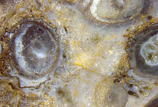
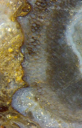 Fig.1
(far left): Ventarura with
and without dark rings on cross-sections.
Fig.1
(far left): Ventarura with
and without dark rings on cross-sections.
Width of
the picture 13.5mm.
Fig.2: Degraded Ventarura, detail
of cross-section on the right of Fig.1, (left to right):
ruggedly shrunk and decayed outer cortex with a layer on the cuticle
partially stained black,
ring-shaped fraction of well-preserved cortex with cell walls partially
stained black, degraded inner cortex, cavity lined with
bluish chalzedony.
The
often conspicuous dark rings (or tubes in 3D) had been interpreted as
sclerenchymatic tissue [1] but
recently re-interpreted as a manifestation of an unexplained
persistence
of some fraction of
middle cortex [2].
(We
apply the terminology used in [1]: The tissue between the central
strand and the epidermis is thought to be subdivided into inner,
middle, and outer cortex.)
Although the aspect of the typical closed ring in the
section on the left in Fig.1 is spoiled by a large dark inner
cortex area
of no relevance concerning the structure of the plant, the
combination of dark ring and rugged outer boundary is strong
evidence of Ventarura.
By queer
coincidence there is a conspicuous dark ring in the section on the
right. It is confusing since it might be
misinterpreted as evidence of Ventarura if
looked at superficially. A closer look reveals that it is only dark
inner cortex, likewise irrelevant as the dark area on the left of this
image. There is evidence of Ventarura,
though less conspicuous: The relevant ring is seen between the
irrelevant
dark
inner cortex fraction and the shrunken outer cortex. This ring
is partially dark or pale, as seen more clearly on the enlarged
detail in Fig.2. Obviously the dark aspect of part of the ring is not
brought about by thick-walled sclerenchyma cells but by some black
deposit, probably of microbial origin, on the
thin walls of rot-resistant cortex cells arranged as a
ring. It has been shown
that the
black deposit had been only loosely connected to the cell wall and may
have flaked
off before silicification [2]. The deposit is probably of the
same type as often
present on the cuticle, where
it may cause the cuticle to peel off and form a curl [2].
No
explanation is proposed here concerning the origin and purpose of the
persistent tissue shaped as a (partial) ring or tube. More questions
arise from a peculiar growth mode whereby the ring or tube became
deformed into a crescent shape (Figs.3,4).
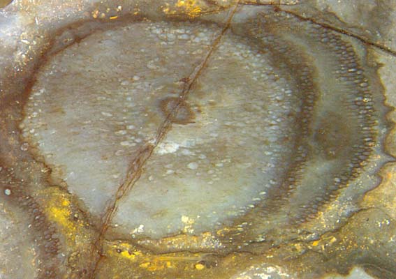
Figs.3,4: Cross-sections of Ventarura shoots
with persistent rings squeezed into crescent shapes by another
shoot grown inside later.
Apparently
a new shoot had grown inside the old one along the outer cortex,
thereby pushing the old tissue aside so evenly that a new circular
outline was formed. The old inner cortex was probably degrading by that
time, offering virtually no resistance. Thus the persistent ring became
crescent-shaped, which implies large deformation during the re-shaping
process. This means that the ring must have been soft, which is another
piece of evidence contradicting the sclerenchyma hypothesis.
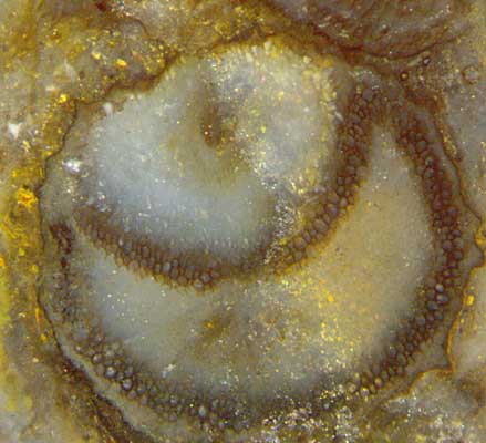
The growth of a new shoot inside the old one is
more advanced in Fig.4 compared to Fig.3. The process can go
so far that the old shoot degrades into
a mere sheath around the new one.
The formation of new shoots in old ones is not rare with the two
zosterophylls
Ventarura
and Trichopherophyton.
It
is not known in which way this process begins. It may also begin in the
inner cortex so that the persistent ring, if there is any, remains
circular as in Fig.5.
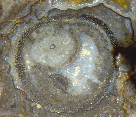
Fig.5: Cross-section of Ventarura
with a small new
shoot inside the persistent ring. Width of the
picture 5.4mm.
Among the cross-sections shown here, Fig.5 is the least confusing one.
With the rugged outline resulting from shrunken and partially vanished
outer
cortex and the well preserved mid-cortex ring, the typical
features of Ventarura
are there.
The
outline is marked by a black deposit on the cuticle. Similar as in
Fig.1, the ring is partially dark or pale. Silicification has preserved
the central strand of irregular shape, a new shoot with own central
strand and circular
cross-section, and some remains of the degraded inner cortex.
Again it is not known where the new shoot came from and where it would
end.
It
has been proposed that new shoots seen inside old ones had grown along
dead plant parts [3] but there are several pieces of contrary evidence.
Hence it remains undecided how to interpret composite plant structures
like those in Figs.3-5 and in Rhynie
Chert News 16.
H.-J. Weiss
2015
[1] C.L.
Powell, D. Edwards, N.H. Trewin:
A new vascular plant from the
Lower Devonian Windyfield chert, Rhynie, NE Scotland.
Trans. Roy. Soc. Edinburgh, Earth Sci.
90(2000 for 1999), 331-349.
[2] H.-J. Weiss:
Rhynie chert - Implications of new finds. EPPC 2014, Padua.
[3] D.
Edwards : Embryophytic sporophytes in the Rhynie and
Windyfield cherts.
Trans. Roy. Soc.
Edinburgh, Earth Sci. 94(2004 for 2003), 397-410.
 |
 |
82 |