Selective decay resistance of soft
tissue in early land plants (2)
 The conspicuous
phenomenon of
small fractions of cortex tissue of
fossil Aglaophyton
and Ventarura
being distinctly seen in Rhynie chert while all
the rest
has vanished has been ascribed to an enigmatic decay resistance in Part
(1). This interpretation contradicts another one proposed
earlier
[1,2], according to which dissolved silica penetrated into a limited
depth below the epidermis of
Aglaophyton, preserving a layer of peripheral
tissue there while the tissue farther below was left to decay. Close
inspection of samples as in Fig.1 provides additional arguments
against an interpretation
according to which the
thickness of the preserved tissue equals the penetration depth of quick
silicification, thereby confirming related doubts raised by A. Channing [3].
The conspicuous
phenomenon of
small fractions of cortex tissue of
fossil Aglaophyton
and Ventarura
being distinctly seen in Rhynie chert while all
the rest
has vanished has been ascribed to an enigmatic decay resistance in Part
(1). This interpretation contradicts another one proposed
earlier
[1,2], according to which dissolved silica penetrated into a limited
depth below the epidermis of
Aglaophyton, preserving a layer of peripheral
tissue there while the tissue farther below was left to decay. Close
inspection of samples as in Fig.1 provides additional arguments
against an interpretation
according to which the
thickness of the preserved tissue equals the penetration depth of quick
silicification, thereby confirming related doubts raised by A. Channing [3].
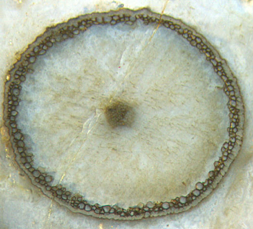
Fig.1: Aglaophyton as
a "hollow
straw" in Rhynie chert: ring of well preserved
cortex cells between the poorly
preserved epidermis and decayed cortex and phloem.
One related argument follows from the fact that the outermost layer of
cells,
the epidermis, is often poorly preserved while a few layers below are
surprisingly well seen. The epidermis is
essentially reduced to a pale structureless ring in Fig.1, except for a
patch on the right, where cell walls
are vaguely visible. So it becomes obvious that the
phenomenon of strongly differential preservation
is more complex than thought at first sight: While most often Aglaophyton is
silicified
rather evenly throughout (see Rhynie
Chert News 60,
Fig.1), less often it
appears
as a hollow straw with poorly
preserved or missing epidermis.
This is
illustrated here with a few more pictures taken from other samples.
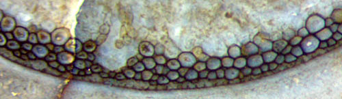
Figs.2,3: Aglaophyton hollow
straw 4mm: well preserved narrow
layer of cortex
cells and poorly preserved epidermis as often seen in Rhynie chert.
Width 1.7mm.
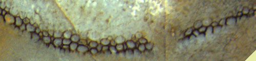
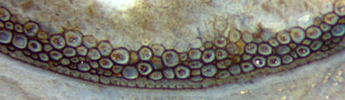
Fig.4: Aglaophyton,
ring of well preserved cortex cells thinning out, epidermis missing,
thus easily confused with Ventarura.
The existence of very narrow and even
discontinuous dark
rings makes
the
stark contrast of small parts of cortex tissue or cells or separate
cell walls even more enigmatic.
The subject does not become less enigmatic when the persistent cortex
rings of Ventarura
are
included into the considerations. They do not seem to differ much from
the rings in Aglaophyton,
except for the fact that usually they are not close to the epidermis.
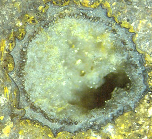
Fig.5: Ventarura
cross-section with persistent cortex ring thinning out towards below
right.
The surface of Ventarura
is
most often shrivelled as a result of shrinkage after decay of the
cortex fraction between the persistent cortex ring and the
epidermis. (The epidermis of Ventarura
has
been found only once, see Rhynie
Chert News 61
).
The
ring can be 5 or more cells thick, and with dark and apparently thick
cell walls it appears strong, hence it has been called sclerenchyma [2]
but
most probably does not deserve such distinction as it is mere cortex
tissue which did not decay for reasons unknown.
Persistent cortex tissue is less readily taken for sclerenchyma in
cases of
- persistent rings thinning out into a string of separate
persistent cells (Fig.5),
- persistent rings without a dark
stain on the cell walls.
(As shown in Rhynie
Chert News 58,
the persistent cell walls are not always stained dark.)
Independent
of the unknown means of protection against decay and of the unknown
purpose thereof, there is another problem involved:
Which
kind of process control, probably
in the live plant, could possibly distribute
the decay resistance among the cells in
such a way that a persistent circle is formed which can even be
discontinuous ? A corresponding question arises in connection with
the rings of fungus-affected cells in Aglaophyton
and Rhynia.
(See Rhynie
Chert News 32).
This may or may not be helpful in the attempt to find an answer.
Finally it can be stated that fossil evidence leads to the
conclusion
that
- the "hollow straw" aspect seen in part of
the Aglaophyton
fossils did not arise from a particular regime of
silica influx,
- the similarity
of the persistent cortex rings in Aglaophyton
and Ventarura
hint at the involvement of a common cause.
The persistent cortex rings seen on cross-sections of Aglaophyton
and Ventarura
offer problems which hopefully may lead to revealing solutions.
Samples: Rh12/91.3: Fig.1; Rh6/38.2: Figs.2,3;
Rh15/35.2: Fig.4; Rh2/143.2: Fig.5;
H.-J.
Weiss
2014
[1]
C.L. Powell, N.H. Trewin, D. Edwards: Palaeoecology and
plant succession in a borehole through the Rhynie cherts, ...
Geological Society, London,
Special Publications 180 (2000), 4 39-457.
[2] www.abdn.ac.uk/rhynie
[3] A. Channing:
Processes and
Environments of Vascular Plant Silicification: Thesis, Chapter
6, Cardiff University, 2001.
 |
 |
66 |