Black shards in decayed tissue
 Clots
of various types and uncertain origin may be found in fossiliferous
cherts. Those which are seen in connection with degraded plant tissue
can often be interpreted as fungus hyphae having
entered into one cell after another [1], having
formed dense tangles there, then partially or wholly consumed the
cell walls. An interpretation is more difficult in the uncommon case
pictured here, where
the clots come with a large variety of sizes
and shapes, often with edges with acute angles, and without any remains
of plant tissue where they could have formed.
Clots
of various types and uncertain origin may be found in fossiliferous
cherts. Those which are seen in connection with degraded plant tissue
can often be interpreted as fungus hyphae having
entered into one cell after another [1], having
formed dense tangles there, then partially or wholly consumed the
cell walls. An interpretation is more difficult in the uncommon case
pictured here, where
the clots come with a large variety of sizes
and shapes, often with edges with acute angles, and without any remains
of plant tissue where they could have formed.
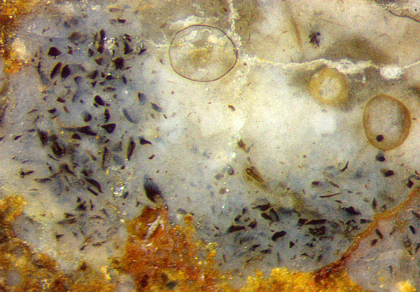
Fig.1: Black shards of various sizes and shapes in bluish
chalzedony replacing Asteroxylon
tissue in Rhynie chert; big chlamydospores of some fungus most
probably not related to the shards. Width of the image 1.7mm.
Closer inspection of the sample sections reveals that the rare
phenomenon covers an area 8mm across where Asteroxylon
tissue had been before but is no more seen after decay, except for
the durable xylem strand.
For comparison, a
picture of a cross-section with some remains of tissue has been taken
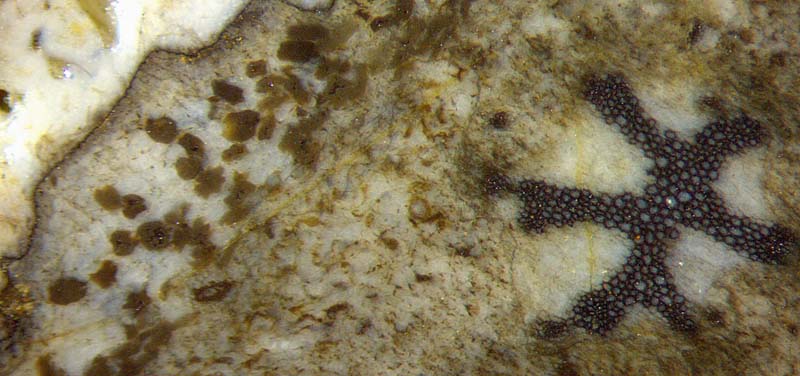
from another sample: Fig.2. There, every dark clot apparently had grown
within a cell, or in two or three adjacent ones, and became angular by
contact with the enclosure. This is the typical growth mode of angular
fungus clots.
Such
interpretation would not apply to the multitude of shapes and sizes in
Fig.1. They could possibly be the result of some microbe entering into
the cells and thriving there to
produce clot matter spreading across the lumen or as layers or crusts
along the cell wall.
After consumption of the cell walls by rot, the odd-shaped replicas or
deposit fragments were left over and became silicified and enclosed in
bluish chalzedony.
Fig.2 (below): Cross-section of Asteroxylon
heavily infested with fungi as usual, with cell-size angular clots
apparently compatible with the vaguely seen remaining tissue. Width
of the image 4mm.
 Asteroxylon
is never seen with well-preserved soft tissue as it is occasionally
seen on Aglaophyton
and Rhynia
sections,
for example. (See Rhynie
Chert News 85.)
While the xylem strand is nearly always well preserved and
makes a spectacular sight, the soft tissue had largely degraded before
silicification, with fungus hyphae running abundantly along the
intercellular spaces, and the cellular structure mostly broken down
(Fig.2). Cells filled with dark matter and the resulting angular clots
are less often seen with Asteroxylon
but more often with other fossil plants. They may be the only left
evidence of cell sizes and shapes from tissues decayed and vanished
shortly after the death of the plant. Their aspect suggests an
interpretation as being due to fungus activity. Fungus chlamydospores
of about 25µm are often seen in decayed Asteroxylon in
large numbers.
Asteroxylon
is never seen with well-preserved soft tissue as it is occasionally
seen on Aglaophyton
and Rhynia
sections,
for example. (See Rhynie
Chert News 85.)
While the xylem strand is nearly always well preserved and
makes a spectacular sight, the soft tissue had largely degraded before
silicification, with fungus hyphae running abundantly along the
intercellular spaces, and the cellular structure mostly broken down
(Fig.2). Cells filled with dark matter and the resulting angular clots
are less often seen with Asteroxylon
but more often with other fossil plants. They may be the only left
evidence of cell sizes and shapes from tissues decayed and vanished
shortly after the death of the plant. Their aspect suggests an
interpretation as being due to fungus activity. Fungus chlamydospores
of about 25µm are often seen in decayed Asteroxylon in
large numbers.
The quite different aspect of the black shards in Fig.1
compared to the angular clots in Fig.2 suggests a similar origin as
that of various black deposits or
coatings, then
left over when their substrates vanished. (See Rhynie
Chert News 83,
87.)
Samples Figs.1,2: found 2001, 2007.
H.-J.
Weiss
2016
[1] M.
Krings,
C.J. Harper, J.F. White, M. Barthel, J. Heinrichs, E.L. Taylor, T.N. Taylor:
Fungi in a Psaronius root mantle from
the
Rotliegend (Asselian, Lower Permian/Cisuralian) of Thuringia,
Rev. Pal. Pal. 239 (2017),
14-30.
 |
 |
103 |