Funny fossil microbes
Fossil microbes are mainly known from stromatolites, where they had
grown as microbial slime and become mineralized layer by layer.
Microbial layers are also found in various cherts formed by
silicification of watery habitats. The filamentous
cyanobacterium Croftalania
for example, forms tufts and wondrous shapes on submerged terrestrial
vegetation
preserved in the Lower Devonian Rhynie chert. Other microbes
which do
not seem to be filamentous, possibly
also cyanobacteria,
can form mm-size emergences occasionally
growing out from abundantly present smooth layers.
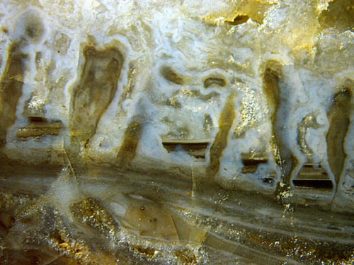
 Figs.1,2:
Microbial layers with upward- growing emergences in Rhynie
chert. Note the levels formed in water between silica gel.
Figs.1,2:
Microbial layers with upward- growing emergences in Rhynie
chert. Note the levels formed in water between silica gel.
Width of either picture 10mm.
Microbial layers as those shown here are seen in a minority of Rhynie
chert samples. Often they are nearly horizontal and only slightly
curved but they
may be folded or broken and tilted after deformation of unknown cause
in a partially silicified state. Distinct
emergences as those seen here are rare.
Conclusions
on the sequence of silicification can
be drawn
from the pictures : The
microbes apparently triggered the early deposition of silica gel which
is
now seen as bluish chalcedony covering layers and emergences. Silica
clusters formed in the water in separate compartments and
settled into suspensions with horizontal surfaces now seen as level faces
separating chalcedony layers of differential aspect. Judging from the
stacks of levels, the deposition proceeded in several stages.
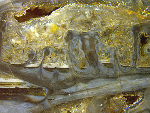
Gel
formation seems to have been more homogeneous in Fig.3, where a tangle
of hyphae of some aquatic fungus is vaguely seen below the stack of
smooth layers
on top. The presence of the fungus is most probably in no way related
to the microbial colonies.
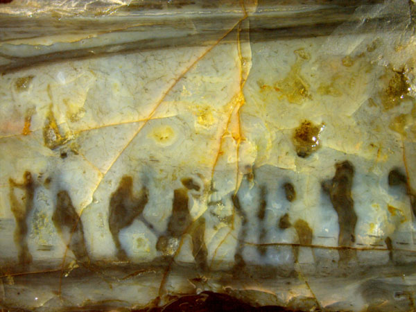
Fig.3 (right): "Crazy performance": emergences between
smooth microbial layers as seen on the raw outside of an old Rhynie
chert
fragment. Width
of the picture 17mm.
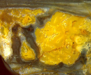 Fig.4 (left):
Microbial formations, same sample as Figs.1-3, later formed chalcedony
stained yellow. Width of the picture 6mm.
Fig.4 (left):
Microbial formations, same sample as Figs.1-3, later formed chalcedony
stained yellow. Width of the picture 6mm.
Fungus
hyphae in the water became coated with clear silica gel which gave rise
to the formation of separate whitish spherulites (poorly seen here in
the yellow area) before the water in
between turned into homogeneous gel and chalcedony. The cause of local
yellow stains is unknown.
As already mentioned, the sequence
of silicification steps can be deduced from the above pictures taken
from one sample. Another sample provides additional evidence (Fig.5): A
smooth layer has broken into pieces
upon emergences from a layer below. This proves that both layer and
emergences were solid
when they got into contact but everything else nearby was fluid. (The
white
dots inside the plant section and elsewhere are tiny spherulites grown
in silica gel.)
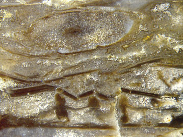 Fig.5: Peculiar assemblage of microbial columns and layers in Rhynie
chert, inclined plant section above. Width of
the picture 8mm.
Fig.5: Peculiar assemblage of microbial columns and layers in Rhynie
chert, inclined plant section above. Width of
the picture 8mm.
 Fig.6: Did the artist anticipate the
existence of microbial formations like those above ?
Fig.6: Did the artist anticipate the
existence of microbial formations like those above ?
Copy from:
www.thescientificcartoonist.com
Annotation 2019: For more microbial emergences see here.
Samples: Figs.1-4: Rh15/63, (0.62kg, obtained from Barron in 2012, here
Part1); Fig.5: Rh9/34.1 (1.55kg, found in 2005, here Part2).
H.-J.
Weiss
2014 2019
 |
 |
67 |