Glades in the nematophyte jungle
Nematophytes are still enigmatic as a whole and in detail as well.
Usually they involve random but rather
even distributions of filamentous tubes occasionally interspersed with
so-called branch knots consisting of a dense tangle of tubes and
supposed to be somehow related to tube
formation [1]. Examples
of such knots, with tubes connected to them, are seen in Rhynie
Chert
News
13, 51,
71.
Clots
of a different aspect but also found between nematophyte tubes deserve
closer inspection. After a first inspection they had been taken for
branch knots in Rhynie
Chert
News 38, 40 but
this must be reconsidered now. The tubes are seen avoiding the
"glades", with only a few
exceptions. There seem to be no tubes obviously starting from there
as they do from branch knots of other nematophytes. Also there
seems to be no inner structure in the glades. The tiny bright dots seen
somewhere are probably glittering crystals, not
sections of tiny tubes.
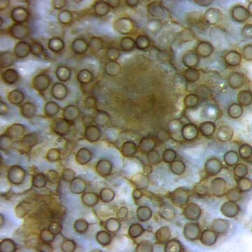
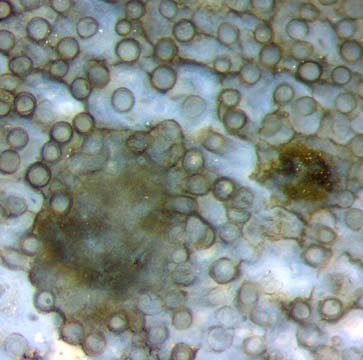
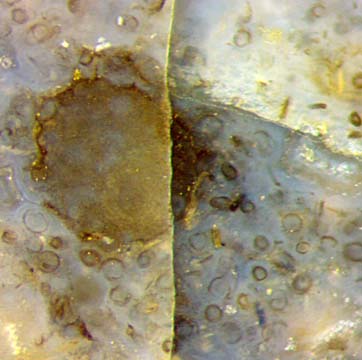
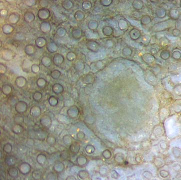
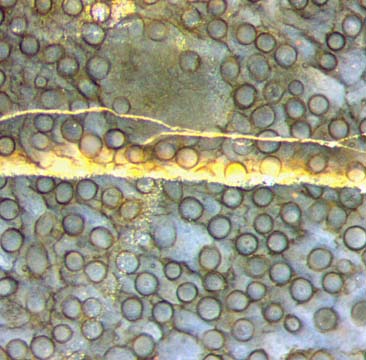
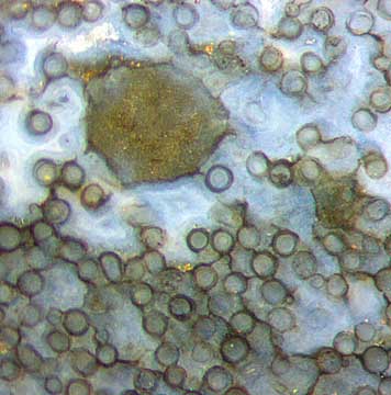
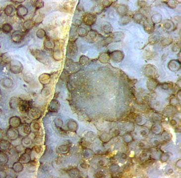
Figs.
1-7: Nematophyte consisting of rather well aligned tubes, mainly
50-60µm across, randomly distributed but apparently avoiding several
rounded "glades" of 0.35-0.5mm seen on the cut face with a frequency of
about 10/cm2.
Fig.3: Partially decayed area.
The size of every image is 1mm2.
Unfortunately,
there is no good lengthwise cut of this nematophyte
available at present so that one cannot be sure whether or not
some
tubes somehow start from the periphery of the glades. The latter
phenomenon is suggested by a few short tube parts with deviating
directions in Fig.2. It is not known why Fig.6 seems to indicate the
contrary: no tube touching the periphery.
Fig.8:
Nematophyte tubes, slightly deranged and separated, thus individually
visible in lateral view. 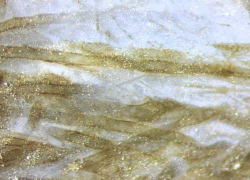
The tiny white dots are due to
the roughness of the raw sample surface. Same scale as above.
Judging from small fracture faces along the
densely spaced
tubes which offer a sideways view, even
a polished
longitudinal section would only offer a
confusing assembly of lines with poor contrast which would
not look like tubes. Individual tubes can be
seen in lateral view if they are displaced
and separated by white chalzedony as in Fig.8.
The small-scale waviness
seen in Fig.8 seems to indicate that the tubes had been rather soft
before silicification, which had also been deduced from observations
in Rhynie
Chert
News 40.
The stronger contrast in Figs. 1-7 compared to Fig.8 does not indicate better
preservation or thick-walled stiff tubes but is simply an optical effect. The thick black wall often
seen on cross-sections may be due to some microbial layer which is
inconspicuous in lateral view. Other microbial sheets
are seen as black lines connecting the tube sections, as in Fig.6.
Finally we are left with the question what to think of the smooth
glades among the tubes which look so much different from the
nematophyte branch knots as we know them.
H.-J. Weiss
2016
[1] www.abdn.ac.uk/rhynie
 |
 |
92 |