A pattern of questionable origin
A conspicuous pattern of radially arranged oblong voids is occasionally
found on plant cross-sections in the Rhynie chert
(Fig. 1). The pattern is well known, for example, from the most
abundant Rhynie plant, Aglaophyton, and has also been found in the
rarest one, Ventarura [1], which belongs to a quite different phylum.
This suggests the existence of a common cause not inherent in the
nature of the plant.
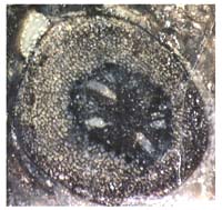
Fig.1: Pattern of voids in a cross
section with otherwise
healthy aspect, probably Rhynia.
Fig.2: Pattern of large voids in a
cross section, probably
of Aglaophyton. Photographs: H. Sahm
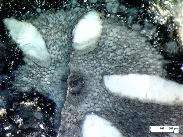
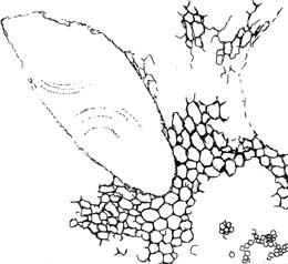 Fig.3: Detail from Fig.2, no indication of decay or rupture
at the apex of the void.
Fig.3: Detail from Fig.2, no indication of decay or rupture
at the apex of the void.
In [2] it is assumed that the void pattern is due
to some decay process resulting in the formation of shrinkage cracks.
This assumption was seemingly confirmed by the observation that the
pattern had been found in decaying axes but not in fresh ones.
Now it appears that the assumption can be refuted by evidence from only
one sample (Fig.2). Although the plant tissue is not completely
preserved on the whole cross section it is seen that the cells near the
alleged shrinkage crack tip are neither decayed nor torn, they look
rather healthy instead (Figs. 2,3). So the
assumption seems justified that the voids had been there
in the living plant *, which requires a not so simple explanation.
The two chalcedony spherulites faintly seen in one of the holes in Fig.
2 could not have been involved in the void formation process: They must
have formed in the already existing void. There is a most likely
explanation based on the following facts:
(1) Fungus infection can cause abnormal growth in
extant plants.
(2) Fungus infection had been identified as a
cause of bloating in Rhynie chert plants [3].
(3) Often Rhynie chert plants are heavily infected
with various fungus species [4].
Hence it can be assumed that the void
pattern was formed in the living plant, with abnormal growth under the
influence of substances released by some fungus hidden in the tissue.
There is room for wild speculation concerning the effect or purpose of
these large voids. They do not seem occupied by the ubiquitous fungus
hyphae
normally seen in the Rhynie chert. If some fungus created them in the
living plant, one might expect that it profited from them in some
obscure way. Also the plant could have profited from the voids as it
could make a larger diameter
with a given amount of tissue and thus become mechanically more
stable. This would be a bizarre type of interaction for mutual benefit.
H.-J. Weiss
2005
[1] C.L.
Powell, D. Edwards, N.H. Trewin : A new
vascular plant from the Lower Devonian Windyfield chert, Rhynie, NE
Scotland.
Trans. Roy. Soc. Edinburgh, Earth Sci.
90(2000 for 1999), 331-349.
[2] www.abdn.ac.uk/rhynie
[3] T.N.
Taylor, W. Remy, H. Hass
: Parasitism in a
400-million-year-old green alga,
Nature 357(1992), 493-494.
[4] T.N. Taylor,
E.L. Taylor :
The Rhynie chert ecosystem: A model for understanding fungal
interactions,
in:
Microbial Endophytes, eds. Ch.W.
Bacon, J.F. White Jr.; Marcel Dekker Inc.
2000.
* Annotation 2014, 2017: For related evidence see Rhynie
Chert News 21, 54, 117.
 |
 |
4 |