Peculiar voids in the tissue of Rhynie
chert plants
Conspicuous
patterns of radially arranged voids on cross-sections of Rhynie chert
plants, like those in Figs.1-5, had been explained as shrinkage cracks
of the decaying tissue [1] before the suspicion arose that fungi
present in the growing plant might somehow have affected growth.
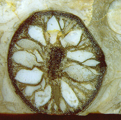
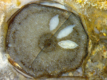
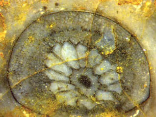
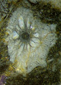
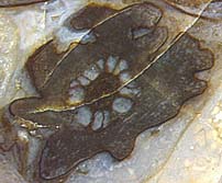
Figs.1-5: Void patterns on Aglaophyton cross-
sections with degraded or well-preserved tissue. All
same scale.
Fig.5 square 6mm.
Extremely
shrunken and kollapsed tissue is obviously present between the large
voids seen in Figs.1,2,3,5 but this does not mean that the formation of
patterns of this kind is merely an effect of degradation. An early
stage of
void formation (Fig.4) suggests that more complex processes must have
been at work. The tissue beside the voids does not look decayed but
deformed as a result of the expanding voids. Apparently there had been
tissue throughout before, and it
became squeezed by the expanding voids while alive. The well-preserved
tissue near the tip of the voids is clearly seen in Rhynie
Chert News
4.
Other
related information comes from enigmatic "twin patterns" where the two
prongs of a forking shoot show essentially the same pattern. Since it
is highly improbable that they developed independently, they must have
been inherited from the base of the fork. (See
Rhynie
Chert News 21, 54.) Some tendency
or inclination towards void formation had
probably become divided along with the forking axis and got into the
branches before
the voids really formed there.
It
is remarkable but confusing that the arrangement of the voids may vary
between chaotic as in Fig.5 and highly pre-determined as in Fig.4.
 As a disturbing
fact, the idea of some fungus being
involved in
the formation of the above patterns is not immediately supported by
fossil evidence. However, supposedly related phenomena as in Fig.6 may
be interpreted in favour of this idea.
As a disturbing
fact, the idea of some fungus being
involved in
the formation of the above patterns is not immediately supported by
fossil evidence. However, supposedly related phenomena as in Fig.6 may
be interpreted in favour of this idea.
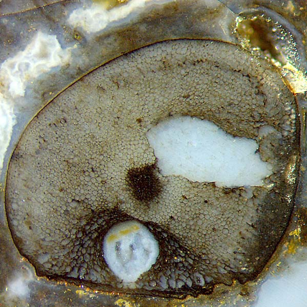
Fig.6: Aglaophyton
cross-section with two types of damage in the live plant: one
hole caused by some unknown herbivore, the other one probably by fungus
activity. Picture 4mm square.
The symmetrical void
in Fig.6 has something in common with the voids in Fig.4: While
expanding, it squeezed the adjacent tissue. The analogy is spoiled,
though, by the observation that the surrounding tissue in Fig.6 is
affected by abnormal growth while the tissue looks normal in Fig.4.
Since abnormal
growth in plants is often due to fungus infection, it can be assumed
that a fungus had been involved here, too. Disregarding
the different
aspect of the expanding cavities in Fig.6 and Fig.4, one may suspect
that both are a result of fungus action.
Chytrid
fungi, which had been present in the Devonian habitat at Rhynie, are
known to be able to bloat plant cells enormously [2]. Hence, the idea
suggests itself that all of the peculiar voids in Figs.1-6 represent
hypertrophy due to fungus action. What remains enigmatic is the way in
which the fungus governs the complex process.
H.-J.
Weiss 2017
[1] www.abdn.ac.uk/rhynie
[2]
T.N.Taylor,
M. Krings, E.L. Taylor: Fossil
Fungi, Elsevier 2015, p.64.
 |
 |
117 |