Voids in the tissue of early land
plants
Conspicuous voids in the tissue of early land plants from the Lower
Devonian,
like those in Fig.1, had been interpreted as shrinkage cracks in the
dead and decaying plant [1]. Closer inspection of
the voids gave rise to doubts. Evidence has been found which indicated
that
the voids were present in the live plant and had been the result of a
growth anomaly, as explained in
Rhynie
Chert News 4,
21.
Since this interpretation is not yet widely known, additional evidence
is presented here. 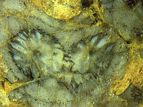
Fig.1: Inclined cut of Aglaophyton
above the forking site of the xylem but below the
forking site of the shoot, with growth anomaly
in
the tissue visible as a void pattern around each prong of the xylem
strand. Width of the picture 8mm.
Evidence for the intricate relationship between early land plants and
fungi is quite common in the Lower Devonian habitat known in a well
silicified state as Rhynie chert.
Cells with dark fills seen on
cross-sections as a concentric ring are due to a phenomenon
known as arbuscular mycorrhiza, which is usually regarded as a kind of
symbiosis [2]. They are seen here as dark dots loosely aligned
at some distance from the circumference (which is
non-circular near the forking site). This shows that some fungus was
present in the live plant.
More evidence for the presence of fungi in live specimens of early land
plants is provided in Figs.2, 3.
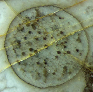 Fig.2: Rhynia, diameter
1.5mm, affected by fungus.
Fig.2: Rhynia, diameter
1.5mm, affected by fungus.
Note that the fungus activity in Fig.2 is not restricted to
a ring of affected cells as often seen in Aglaophyton
and less often in Rhynia.
Although the tissue is affected throughout, it is well preserved so
that one can assume that this fungus, too, grew in the live plant
without causing damage. (See also Rhynie
Chert News 32.)
Another type of fungus invasion is seen in Fig.3, where hyphae had
grown in an essentially parallel way along the shoot of Aglaophyton where
they are faintly seen now. Here the central strand had been above the
cut plane and hence cut away. For reasons unknown, most of the tissue
had
vanished so that the hyphae are better visible here than in the
presence of tissue. If they had grown in the dead and
decaying plant, they would more probably have grown in any direction,
hence the parallel alignment seems to indicate
that they, too, grew in the live plant.
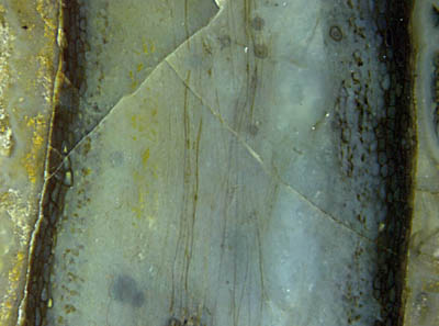
Fig.3 (right): Aglaophyton
shoot, diameter 4.5mm, apparently hollow, with faintly seen hyphae
arranged lengthwise.
The
abundance of fungi met in the early land plants [2], be they symbionts
or
parasites, suggest that they may be responsible for the big and small
holes in the tissue which cannot be ascribed to shrinkage or herbivory.
This idea is supported by the rare cases of two roughly
mirror-symmetric void patterns ("twin patterns"). Previously presented
evidence has been based on
separate "twins"
but Fig.1 shows the twin patterns before separation. They
could hardly form independently but can be explained as having been
transferred from the base of a forking shoot into the growing
prongs of the
fork. (This applies also to Rhynie
Chert News 55,
Fig.1.) Hence, the voids are very probably due
to misguided growth
triggered by substances released by a fungus, and discussing void
formation in live plants as a subject of taphonomy as done in [1] is
severely erroneous.
Now that an explanation of the voids, contrary
to established views and with the help of twin patterns, has been
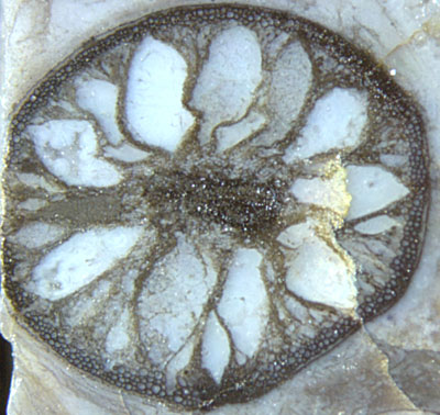
proposed here, a particularly conspicuous
flower-shaped pattern is shown here just for its beauty (Fig.4).
This seems to be an extreme case of misguided growth but it is also
thinkable that the pattern is the result of more than one process
stage. Judging from the aspect of the tissue along the circumference,
the plant could well have been alive while its interior was reduced to
several strings keeping the central strand suspended in the
centre.
Fig.4: Aglaophyton
cross-section, 5mm, with conspicuous void
pattern.
H.-J.
Weiss
2013
[1] www.abdn.ac.uk/rhynie
[2] T.N. Taylor,
E.L. Taylor :
The Rhynie chert ecosystem: A model for understanding fungal
interactions,
in:
Microbial Endophytes, eds. Ch.W.
Bacon, J.F. White Jr.; Marcel Dekker Inc.
2000.
 |
 |
54 |