Black and white stains in
silicified wood
Small
fragments of coniferous-type fossil wood are of little interest in
palaeobotany unless they provide evidence of processes or
structures other than mere silicification of plain wood. The central
pith, which clearly differs from the "proper" wood, is seldom preserved
and
therefore deserves attention. As a remarkable fact also seen here, the
pith cell size decreases near the boundary towards
the wood. In the course of silicification, part of the silica
gel had formed crystallites with
sizes exceeding the light wavelength so that they reflect the light
and, if in large numbers, appear snow-white. In
this way, parts of the pith and the wood, including the boundary region
between pith and wood, had become surprisingly well visible.
The dark spot
of destroyed tissue between the white cells in Fig.2 suggests microbial
activity as a possible cause of this crystallisation.
Not understood is the cause of the squeezed aspect of
part of the cells along the boundary, notably in Fig.1.
This sample reveals also another phenomenon which
is quite the opposite of whitening: It is the blackening of cell
walls, which seems to be another
indication of the involvement of microbes. Individual cells may differ
clearly from their surroundings by blackened walls.
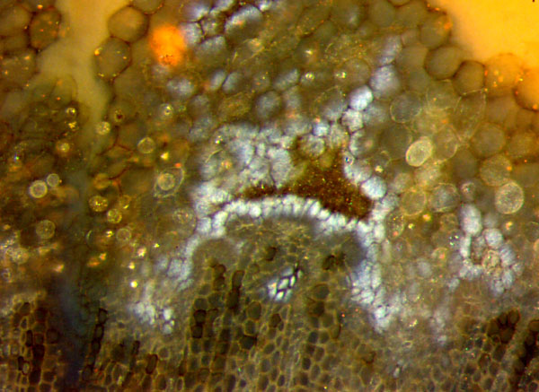
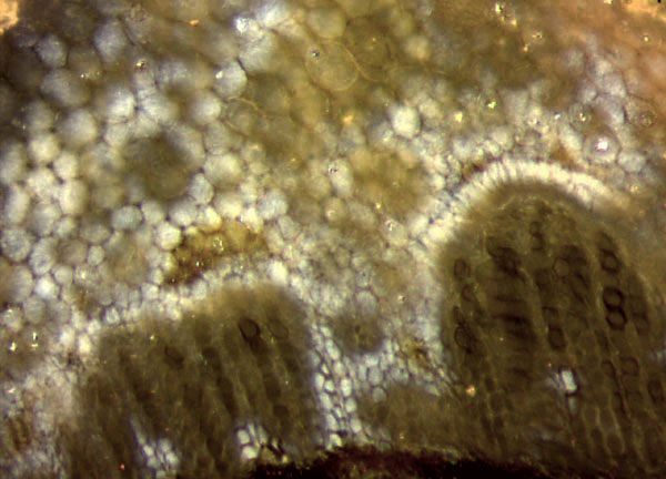
Figs.1,2:
Coniferous-type wood with pith, whitened cells, and blackened cell
walls;
Lower Permian.
Height of the images 1mm.
Pictures taken from the two cut halves of Sample Kc/24 found in the 90s
at Kleincarsdorf
near Kreischa,
Doehlen basin, Saxony.
From the observational fact that the black stain occasionally does
not cover the whole cell wall but only part of it as seen with some
cells in Fig.2, one may conclude that it is due to the
spreading
growth of some microbe.
Cell walls with black coatings can provide the
illusion of cells with dark fill, as in Fossil Wood News 21
,
for example. There can be more than one cause for the dark appearence
of cross-sections of individual cells. Varying
shades of gray along the tracheids in Fossil Wood News 35
(there Fig.2), seem to be due to microbes which had lived in the cell
lumina before silicification.
The
conspicuous lonely cell with white lumen and black wall in Fig.1 serves
as evidence that the processes of blackening and whitening did
not
mutually interfere. The same applies to the white spot spreading across
a few black cell walls in Fig.2.
Thin black coatings on surfaces and cell walls of the early land plants
in the Rhynie
chert are
also due to microbial activity. (See Rhynie
Chert News 83, 87.)
H.-J.
Weiss 2019
|

|
 36 36 |