An uncommon variety of Rhynie
chert
 Judging
from numerous own finds and from samples seen in collections
and as images in publications, the vast majority of them are more or
less dull dark if looked at from afar and reveal their rich
detail, if there is, under the microscope, preferably with transparent
light [1,2]. Those with much different aspect
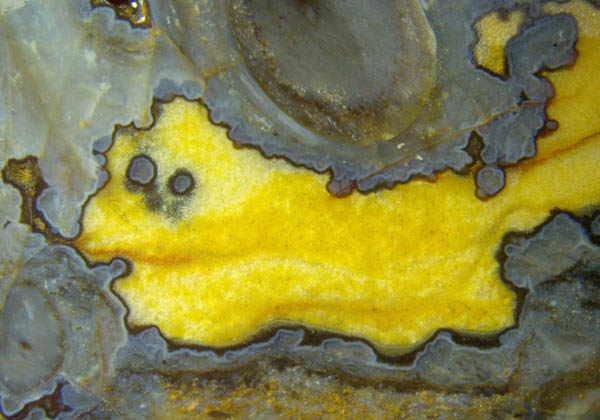
are rare, as the one described here with bright yellow fills in former
cavities in the chalcedony: Fig.1. (The bright yellow has turned dull
ochre with time on
and near the surface of this old chert layer fragment.)
Judging
from numerous own finds and from samples seen in collections
and as images in publications, the vast majority of them are more or
less dull dark if looked at from afar and reveal their rich
detail, if there is, under the microscope, preferably with transparent
light [1,2]. Those with much different aspect
are rare, as the one described here with bright yellow fills in former
cavities in the chalcedony: Fig.1. (The bright yellow has turned dull
ochre with time on
and near the surface of this old chert layer fragment.)
Fig.1: Rhynie chert, former cavity filled with a granular
quartz
deposit of yellow aspect, with dark lining, probably of microbial
origin, 40µm. Width of the image 1cm.
What has been called here a lining of the cavity can as well
be called a coating
on the
surface of the bluish chalcedony. As a probable scenario, inundated
plant
debris and other substrates in silica-rich water triggered the
deposition of
silica gel with irregular surface and bulging protrusions.
Apparently the
deposition ended when most of the dissolved silica was used up.
Subsequently a layer
of microbial slime grew on the gel surface and became silicified later,
with microbes enclosed (Figs.2-4).
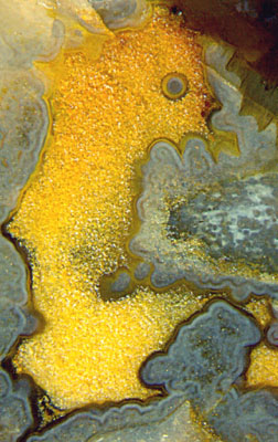
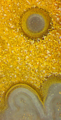
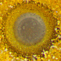
Figs.2-4:
Similar as Fig.1, with dark or
pale lining, with
tiny black dots
(2µm) seen scattered around the boundary of the gray area in the "eye".
Fig.4: 0.4mm.
Obviously the dark aspect is not an intrinsic property of the coating
or lining of about 40µm thickness.
It seems to be a secondary phenomenon, in the present case caused by a
black stain taken on by a smaller or larger fraction of the particles
which the layer seems to contain, producing shades varying
from pale brown to black.
Individual black dots are often seen a few µm above the layer, which
probably indicates that their position had been fixed by organic gel
before silicification.
There seems to be a connection with similar coatings and deposits
which
are conspicuous only if stained dark, as often seen with parts of
tissue of Aglaophyton
and Ventarura.
After
nearly all of the initially supplied dissolved silica had been used up
in gel formation, and the gels in the bulk and the coatings and the
silicified plants had become such solid matter on their path of
transformation into chalcedony that cracks had been able to run
through as if the whole were brittle, slow diffusional influx of silica
caused the growth of what is now
seen as a deposit of quartz crystals of yellow aspect, filling cracks
and cavities alike (Fig.5).
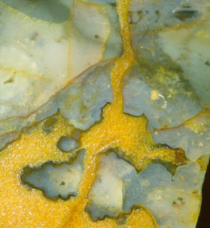
Fig.5
(left): Wide crack running through silica gel and crossing a cavity
with dark lining. Note the absence of any lining on the crack faces.
The small black level fill above right indicates the up-direction
during silicification. Width of the image 5mm.
The
former cavities of unusual aspect due to their dark lining and yellow
fill in Figs.1-7,10 differ much from the more common former cavities in
the chert which most often can be traced back to gas bubbles which got
stuck among submerged plant debris or microbial mats in silica-rich
water (Figs.6,8,10). Apparently the bubbles remained empty while
everything around turned into silica gel. Later the gas escaped by
diffusion and water entered likewise. Usually, fungus hyphae thrived in
the water-filled cavities, now seen
coated with bluish chalcedony (and quartz, Fig.8), later often
completely enclosed, as in Fig.10.
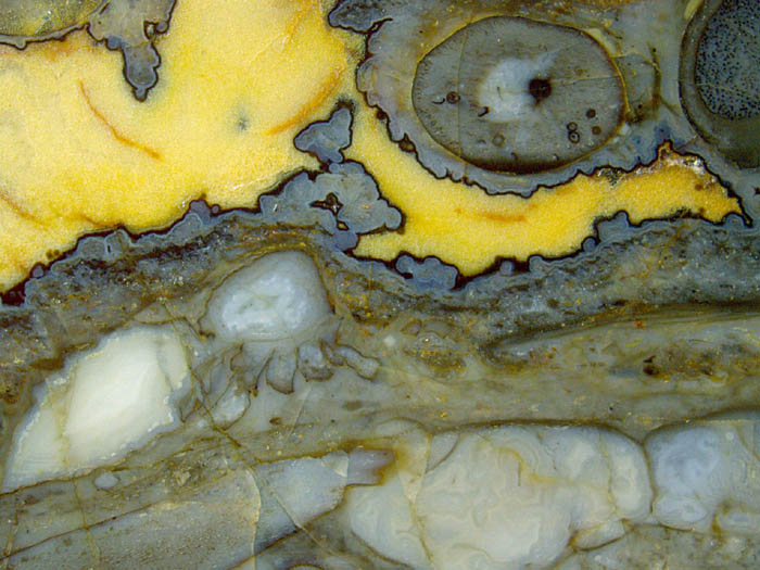
Fig.6 (right):
Chert with former cavities of various origin, now filled with
chalcedony or quartz. Note the lengthwise cut nearly along the symmetry
plane of a complete
trigonotarbid or its moult, rare big species (on the left). Width of
the image 17mm.
Several types of former cavities can be distinguished in
Fig.6:
- spaces without silica gel, water-filled, now
filled with quartz, here yellow,
- gas bubbles in swamp matter (below right), now
filled with bluish chalcedony,
- the empty hulk of a big terrestrial arachnid [2],
with agate-like fill,
- a void in Aglaophyton
(above), now filled with bluish chalcedony.
Another type of former cavity is seen in Fig.7:
- crescent-shaped narrow cavities due to shrinkage
of embedded plant shoots,
now filled with
chalcedony and quartz.
Note
that there are two characteristic features of the first-mentioned type
of
cavity: distinct linings, absence of hyphae. The latter
fact seems
to indicate that either there had never been hyphae because all organic
matter
was sealed within solid gel and thus inaccessible to fungi or else if
there
had been hyphae, influx of dissolved silica was so slow and late that
the
hyphae did not become coated with gel and hence vanished before the
cavity
gradually got filled with quartz.
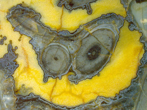
Fig.7:
Two sections of Aglaophyton
once enclosed
in silica gel, then slightly shrunken and detached before
silicification. Width
of the image 17mm.
Obviously the plants in Fig.7
have shrunk away
from their enclosure of silica gel, which indicates delayed
silicification inside, probably due to the presence of the cuticle, so
that the tissue was degrading and shrinking even after the enclosure
had become rather solid.
Several tiny details on the sections are due to fungi in the live plant.
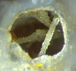
Fig.8: Cavity with coated fungus hyphae, thickness 0.2mm.
The hyphae of
aquatic fungi are common in the Rhynie chert, notably in places where
silicification was so slow that free water persisted for some
time. Gas
bubbles which later
became filled with water were suitable places for
growing hyphae. Much later the cavities would either become filled with
chalcedony or quartz as
in Fig.6 (below) or the water would vanish after the once delicate
hyphae had been coated and turned into rods
crossing the now empty cavity, which makes an impressive sight (Fig.8).
The
presence of fungi
in this Lower Devonian habitat reveals itself in the
Rhynie chert also in other ways. Cells with dark fill, loosely
aligned as a concentric ring on cross-sections (Fig.7), are a
characteristic feature of the symbiotic fungus Glomites rhyniensis
[3].
Quite different fungus parts are the vesicles summarily called
chlamydospores. Some of them are seen on the plant section in
Fig.6. Occasionally
something is attached to them which looks like the collapsed remains of
an older vesicle, and rather seldom the attached object and the
connecting tube are still in
good shape (Fig.9, see Addendum below).
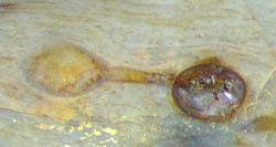
Fig.9 (right): Fungus chlamydospore inside Aglaophyton with
attached object of similar size. Width of the combined object 0.9mm.
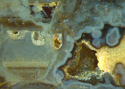
Fig.10
(left): Former cavities of different origin,
with enigmatic partial fills. Width of the image 4.5mm.
Fungus
hyphae grown in water-filled cavities are also seen in Fig.10, although
less spectacular than in Fig.8. Stacks of level layers are not rare
in the Rhynie chert. The layers below left had
obviously been deposited in a globular water-filled former bubble
after a few hyphae had grown there and had got a transparent coating of
silica gel, now seen as bluish chalcedony looking like wormholes in the
layered deposit below. A straight hypha is faintly seen below the
pocket of granular quartz on
the left. Bluish inclusions in the dark level fills of other former
cavities
above left are coated hyphae, too.
What is seen on the right half of Fig.10 is of the same
type as the quartz-filled cavities in Figs.1-7 except for the fact that
it is not quite filled and never had been. There are more of such
cavities partially filled with quartz grains which did not assemble at
the bottom, which is an unexplained phenomenon.
Other uncommon details are mentioned here only briefly:
There are long
slender bristles (not seen here), thickness about 2µm, on the legs of the rare big trigonotarbid species in
Fig.6.
The spore capsule of Aglaophyton,
part of which is seen in Fig.6 above right, lacks the typical feature,
the palisade wall aspect of the outer capsule
wall. This may be due to the fact that it is a juvenile sporangium,
judging from the observation that all spores are still in
tetrads, a stage less often seen preserved in chert.
Obviously this small sample of
Rhynie chert, conspicuous for its cavities
with yellow fill and distinct dark lining,
offers an unexpectedly rich trove of detail, more or
less understood or still enigmatic.
Sample: Rh7/10 (0.23kg), found in 2003
by Sieglinde
Weiss
near Milton of Noth. The pictures have been taken
with incident light.
Addendum
Fungus formations as in Fig.9 have been thoroughly described in
[4] as "acaulosporoid glomeromycotan spores" forming "spore-saccule
complexes". The spore is thought to develop sideways on the tail of a
saccule. This means that the visual impressions from Fig.9 above and
from Fig.5 in Rhynie
Chert News 55
, which suggest a straight connection by a thick tube, should
be regarded as mere illusions. Apparently
the deeper question why a big spore develops from an equally big
saccule on a thin hypha is dealt with in the ample fungus literature
referred to in [4].
H.-J. Weiss
2014 2020
[1] N.H. Trewin, C.M.
Rice (eds.): The Rhynie hot-spring system:
Geology, biota, and mineralisation.
Trans. Roy. Soc.
Edinburgh, Earth Sci.
94(2004 for 2003) Part4, 283-529.
[2] H.
Kerp, H. Hass : De Onder-Devonische Rhynie Chert,
Grondboor & Hamer 58(2004),
33-51.
[3] T.N.
Taylor et al.: Fossil arbuscular mycorrhizae from
the Early Devonian,
Mycologia 87(1995), 560-73.
[4] N.
Dotzler, Ch. Walker, M.
Krings, H. Hass, H. Kerp, T.N.
Taylor, R. Agerer:
Acaulosporoid
glomeromycotan spores with a germination shield from ... Rhynie chert.
Mycol. Progress (2009) 8,
9-18.
 |
 |
64 |