Venation pattern – a distinct feature
of Psaronius tree
fern pinnules
An introductory remark is appropriate here to avoid confusion since
there
are several valid names for different parts of the same fossil plant:
The conspicuous
trunks with their intricate internal structure are known as Psaronius
*.
Their
foliage has got the name Pecopteris
when preserved as a compression fossil but
Scolecopteris
(which literally means “maggot
fern”) when preserved 3-dimensionally with sporangia present.
The ambiguities introduced in this way shall not concern us here.
With increasing amounts of "maggot stones" from Döhlen basin (Lower
Permian)
becoming available since 1993 it has become apparent that some features
of the “maggot fern” vary within wide
bounds: The pinnules may be nearly straight or curved up to
half-circular
shape. Their margins may be nearly smooth or beset with more or less
long and tapering fringes. The number of sporangia fused into a
synangium may
vary from 3 to 6 or rarely 7, and even for a given number of sporangia
per synangium their pattern of arrangement may
differ on one pinnule [1].
It will be difficult to find out to which degree such variability is
due to environmental factors (sun or shade, more or less wet habitat,
salinity of swamp water), age
(juvenile, adult, wilting), etc., and which are inherited features.
Finding out this might give clues concerning the response to a changing
environment, or it might serve as a basis for differentiation between
various strains or species.
There are at least two features which are most likely inherited: (1)
the presence or
absence of forking lateral veins on the pinnules and (2) the angle
between the lateral veins and the pinnule axis. According to Millay
[2], forking veins are absent in Sc.
elegans
and several related species. Scolecopteris
specimens with forking veins
are not rare among those recently
found in Döhlen basin, which is incompatible with the claim in [3,6]
that all recent finds belong to Scolecopteris
elegans.
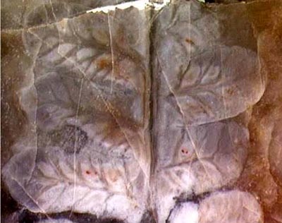
Fig. 1: Pinna of Scolecopteris
with both forking and simple veins on the pinnules.
Width of the picture 6mm.
Although
much more structure information can be preserved in chert compared to
coalified compressions, the vein pattern of the chert specimens is less
conspicuous because the fronds and their parts are usually not lying
flat in the chert but in tilted and distorted positions. Even in the
rare cases where pinnules form a surface relief, the vein pattern is
usually seen less
distinctly than on the often larger frond parts found as
compression fossils. However, there are chert samples with
largely
decayed plant matter where virtually nothing is left but a few forking
veins revealing the presence of fern
foliage.
When trying to measure the angle at which the
veins branch off, one encounters more difficulties: The
pinnule shape implies that the veins are not
curves in a plane but in space, and their curvature tends to vary
shortly after branching off from the midvein. In order to agree upon a
simple procedure of angle measurement, one may choose the tangent to
the vein at a point halfway to the margin, and look at that tangent in
top view, which means project it onto the plane of the pinnule. The
pinnule, however, is usually not plane, hence one has to take the
tangent plane to the pinnule at the position of branching-off for a
reference plane.
This is about the same what one would perhaps intuitively do without
such set
of rules but it may be useful to be aware that other rules would
produce other values of the angle.
Independent
of the details of measurement, a careful look at the veining angles
and other features reveals that the literature
on Scolecopteris
is fraught with a disturbing lot of
inconsistencies waiting to be removed.
The venations of Fig.2 and Fig.3
differ significantly. By taking the intuitive approach, one finds
angles around 60° in Fig.2 and 45° in Fig.3. Hence, if the angle is
typically 60° for Sc.
elegans as claimed in [3], the pinnules in Fig.2 may be Sc. elegans but
those in Fig.3 are certainly not. (This is also supported by their
length of 7mm since the length of Sc.
elegans
pinnules is 4-5mm [3].) If Fig.3 were Sc. elegans as
claimed in [3], Tafel 2, the 60°-rule would not
be valid.
What is presented as Sc.
elegans
in [4], Tafel VII, and [3], Tafel 5, shows veining angles of 45° and
other features differing strongly from those of the type specimen
(lectotype) in [3], Tafel 1. 45°-venation on alleged Sc.
elegans is also seen in
[5], Fig.6. (There, "Pecopteris
elegans" in the captions of Figs.3,4,6-10 is a mistake.)
The pinna in Fig.1 differs from Sc.
elegans not only by its forking veins but also by angles
smaller than 60°.
When the chert sample with the pinnules pictured in Permian Chert
News 1,
Fig.1, seen on the raw surface in
side view, was recently cut, it
revealed pinnules with 45°-venation
on the cut face, which is not compatible with its assignment
to Sc.
elegans in
[3].
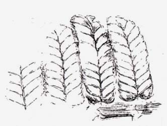
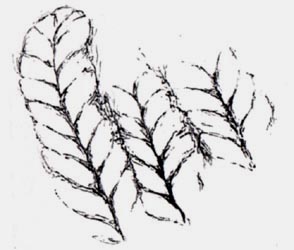 Figs.2,3: Rare examples of Scolecopteris
pinnules distinctly seen as reliefs on the surface of a chert layer.
Height of the pictures 7mm.
Figs.2,3: Rare examples of Scolecopteris
pinnules distinctly seen as reliefs on the surface of a chert layer.
Height of the pictures 7mm.
It requires some experience to reveal a vein pattern like the one
in Fig.1 by
carefully choosing the cut plane of the sample in such a way that as
many pinnules as possible are seen in top view. In this
connection it is worth mentioning that Gert Müller
(Dresden) [7], who found and cut the sample of
Fig.1, was the first one to
find more “maggot stones” after nearly a century of no finds, thus
essentially contributing to the recent upsurge of interest in the
subject.
Finally it appears that distinct features of the vein
pattern,
as forking veins and angles near 45°, provide strong arguments against
the interpretation of all recent finds of Scolecopteris in
Döhlen basin as Sc.
elegans,
as claimed in [3,6]. This is supported by other features,
as the presence or absence of hairs, long or short pedicels
of
synangia,
enclosed or exposed synangia,
and others.
Samples: Old fragments of Lower Permian chert
with more or less rounded
edges, found among younger fluviatile
deposits in Döhlen basin near Dresden, Saxony, Germany.
Fig.1: Found by Gert
Müller
(Dresden) [7] at the type locality of Sc.
elegans near
Kleinnaundorf (1985), photograph
by H. Sahm.
Fig.2: Sample W/3.2, own find (1992), Wilmsdorf,
golf course.
Fig.3: Sample H2/35.2, own find (1993),
Hänichen.
* Uncommonly big and well-preserved specimens
of Psaronius,
the stem of several "maggot fern" species, are on display
at the Naturkunde Museum Chemnitz.
H.-J. Weiss
2011
[1] H.-J.
Weiss: Beobachtungen zur Variabilität
der Synangien des Madenfarns. Veröff. Museum f. Naturkunde Chemnitz
25(2002), 57-62.
[2] M.A. Millay:
Study of paleozoic marattialeans. A monograph of the American species
of Scolecopteris.
Palaeontographica B169(1979), 1-69.
[3]
M.
Barthel, W. Reichel, H.-J. Weiss : "Madensteine" in
Sachsen. Abhandl. Staatl. Mus. Mineral.
Geol. Dresden 41(1995), 117-135.
[4] M.
Barthel : Pecopteris-Arten
E.F. von Schlotheims aus
Typuslokalitäten in der DDR. Schriftenreihe geol. Wiss. Berlin
16(1980), 275-304.
[5] M.
Barthel, H.-J. Weiss: Xeromorphe Baumfarne im Rotliegend
Sachsens. Veröff. Museum f. Naturkunde Chemnitz
20(1997).
[6] M. Barthel:
The maggot
stones from Windberg ridge, Germany.
in: U. Dernbach, W.D.
Tidwell: Secrets
of Petrified Plants. D'ORO Publ. 2002. p65-77.
[7] G. Müller:
private communication.
|
 |
 2 2 |