A look at Devonian fungi
Fossil fungi are rare and such an exotic subject even for
palaeobotanists that fossil collectors may be led to the assumption
that it is no use looking for them. The enigmatic Prototaxites from
the
Silurian and Devonian, which comes in sizes of big tree trunks, has
recently been interpreted by some as an enormous fungus or lichen.
Putting aside that problematic fossil, not much is left
that looks like Palaeozoic fungi, with a remarkable exception: Fungi
are quite abundant in the Rhynie chert, which is famous for
the
good preservation of a waterlogged habitat from the Lower
Devonian. Well, one needs a microscope to find them and to see in which
way they had affected the living and decaying plants. The delicate
hyphae may be more easily spotted if they extend
across a cavity, strengthened by a
coating of chalcedony or quartz crystals (Fig.1).

Fig.1: Inclined cut of a hollow straw of Aglaophyton with
dark wall of preserved tissue and coated
fungus hyphae traversing the cavity. Width of the image 4mm.
The
hyphae of the aquatic fungus had grown when the cavity was filled with
water. Later they got a thick coating of chalcedony topped with quartz
crystals. Note the hypha seen in full length through the translucent
coating and others seen as tiny dark dots on cross-sections.
Incidentally,
other manifestations of the presence of fungi are seen within 3cm on
the same cut
face of the small chert sample of 120g where the coated hyphae have
been found. Rather common are the
thick-walled globules of about 0.35mm diameter,
usually called chlamydospores, in decaying Aglaophyton in
Fig.2 below left.
Another
fungus species is seen on the odd-shaped section in the centre. Its
chlamydospores are comparatively tiny, with diameters of 0.05mm.
Although the odd shape of the section is not important here, it may be
explained: It is an inclined cut of Aglaophyton,
non-cylindrical near a forking site, possibly slightly
squeezed in addition to the natural asymmetry. Also not related to the
fungus is the conspicuous crack on the left, formed in a state of
advanced
silicification when the response to strain was no more dominated by the
plant tissue but by the mechanical parameters
of the homogeneous silica gel. Later it became filled with a
yellow deposit.
A quite different phenomenon is seen on the right of Fig.2. With the
exception of the conducting strand, the tissue seen
on the cross-section
is obviousy degraded in a particular way:
Small voids have formed in the cortex which are so evenly distributed
on
average that they do not look like a result of random rot. They rather
resemble the "flower-shaped" void patterns, a growth anomaly
probably controlled
by some fungus.
The presence of a fungus in the cortex tissue in (Fig.3)
is revealed by a globule,
slightly shrunken now,
which seems to fit into the mesh. It is not known
whether or
not it belongs to the fungus responsible for the patterning.
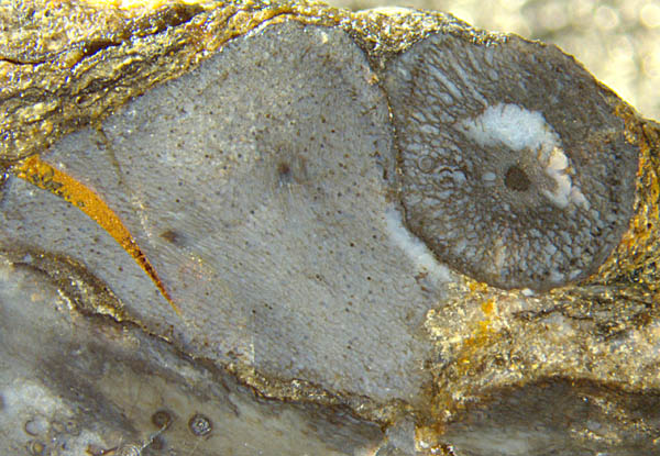
Fig.2 (left): Aglaophyton
sections of different aspect, with at least
four different
manifestations of fungus activity.
Width of the image 8.6mm.
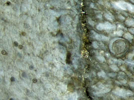
Fig.3: Detail of Fig.2, boundary separating Aglaophyton axes
affected by fungi in different ways. Width of
the image 1.4mm.
The fungus which reveals its presence by a characteristic
dark fill of cells in a loose row, some of
them collapsed, left of the vertical boundary in
Fig.3, is easily
recognized, with the help of [1], as the
symbiotic fungus Glomites
rhyniensis,
which spreads in the live plant. The dark fills consist of so-called
arbuscules, dense tangles of very thin branched hyphae engaged in
symbiosis. The globose objects on the left, diameter 45-55µm,
apparently correspond to what is called spores in [1] although their
function is said to be uncertain. Those spores differ slighty from the
present globules as they are "nearly elongate to globose and range from
50-80µm" [1].
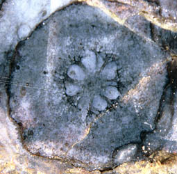
The
suggestion that what is seen on the right of Fig.2 is a growth anomaly
implies
that another fungus species is able to occupy the live plant. This may
be illustrated by images of "flower-shaped"
void patterns from other samples, one
of which is shown in Fig.4. An
interpretation of similar voids as being due to mere shrinkage of
decaying tissue
[2] can
be ruled out by the observation that there is healthy tissue adjoining
to the voids. Other fossil evidence most probably indicating the
presence of fungi in live plants is presented in Rhynie
Chert News 54.
Fig.4 (right): "Flower-shaped"
void pattern in Aglaophyton suggesting
its formation by
anomalous growth controlled by
some fungus. Width of the section 3mm.
It
appears that fossil fungi from the Palaeozoic deserve due attention
since they had been essential for the colonisation of the land
by plants [3], and scientific work seems to cover only part of the intricate
but fascinating
subject [4,5].
(Some of the size data in [4] and other publications on Devonian fungi
are mutually inconsistent and hence erroneous. See chapter Errors.)
Samples: Rh5/9 (0.12kg), found in 2002. Part 1: Figs.1-3.
Rh9/73 (0.33kg), found in 2004. Part 2: Fig.4.
H.-J.
Weiss
2014
2020
[1] T.N.
Taylor et al.: Fossil arbuscular mycorrhizae from the
Early Devonian,
Mycologia 87(1995), 560-73.
[2] www.abdn.ac.uk/rhynie
[3] T.N.
Taylor, J.M. Osborn: The importance of
fungi in shaping the paleoecosystem.
Rev. Palaeobot. Palyn.
90(1996), 249-262.
[4] H. Hass, T.N.
Taylor, W. Remy: Fungi from the
Lower Devonian Rhynie chert.
Amer. J. Botany 81(1994),
29-37.
[5] T.N. Taylor,
E.L.Taylor, M. Krings: Paleobotany, Elsevier 2009.
 |
 |
63 |