Nematoplexus
with controversial aspects
 The enigmatic organism Nematoplexus
had become known from apparently only one sample of Rhynie chert
collected
decades ago [1]. Since 2003, a few more samples have provided evidence
that
Nematoplexus is
even more enigmatic than previously thought [2]. The new evidence gave
also
rise to doubts concerning the dark lumps called "branch-knots". They
are supposed to produce the tubes (Fig.1) and, according
to [3], to
consist of "very tightly coiled ... tubes
showing repeated and closely spaced branching". The latter part of this
quotation
refers to [1] (there Plate 1, Figs.7,10) with tubes of about 2...8µm
diameter, hence it does not concern the coiled tubes of 9...15µm seen
here.
The first part of this quotation, "very
tightly coiled", is an unsubstantiated addition since nothing like this
is seen in [1] or in the own samples.
The enigmatic organism Nematoplexus
had become known from apparently only one sample of Rhynie chert
collected
decades ago [1]. Since 2003, a few more samples have provided evidence
that
Nematoplexus is
even more enigmatic than previously thought [2]. The new evidence gave
also
rise to doubts concerning the dark lumps called "branch-knots". They
are supposed to produce the tubes (Fig.1) and, according
to [3], to
consist of "very tightly coiled ... tubes
showing repeated and closely spaced branching". The latter part of this
quotation
refers to [1] (there Plate 1, Figs.7,10) with tubes of about 2...8µm
diameter, hence it does not concern the coiled tubes of 9...15µm seen
here.
The first part of this quotation, "very
tightly coiled", is an unsubstantiated addition since nothing like this
is seen in [1] or in the own samples.
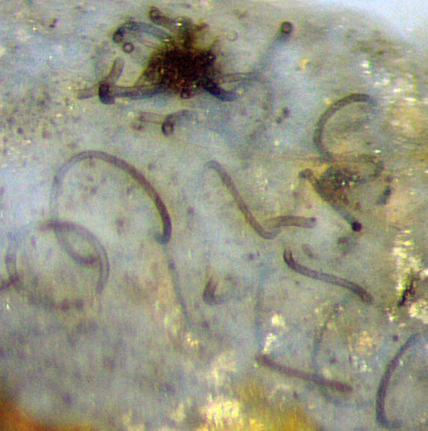
Fig.1:
Nematoplexus
as seen on the raw surface of a Rhynie chert sample, with two dark
lumps called branch knots among spiralling tubes. Image width 0.6mm.
For separate images of the lumps taken by Gerd Schmahl with
higher resolution, other illumination,
and other orientation of the sample, see Figs.2,3.
It would be hard to imagine that the tubes seen emerging from the dark
lump in Fig.2 had lain "tightly coiled" and branched inside.
The tiny lump in Fig.3 is less confusing than the bigger ones in Fig.2
or elsewhere
in this sample. It shows two spiralling tubes emerging from the lump.
The right one is seen bending towards the observer, then
turning into the depth, vanishing from sight, then emerging and
vanishing again. Hence, nearly two turns with right-hand thread are
seen. With the uneven surface of this
sample and other things
precluding a proper visual impression, the tube
on the left may provide the illusion of left-hand thread but comparison
with the differently oriented sample in Fig.1 shows that the thread
is right-handed as expected. A third tube in Fig.3 is
seen as a cross-section on the raw sample surface.
Again it
appears that there is not much room for coiling and branching of the
three tubes in the small "branch-knot", hence the tubes must have
formed there somehow more immediately. Their way of formation remains
obscure but there are indications from other samples that
they may form
at or near the surface of the lump. (See Rhynie
Chert News 135.)
Anyway, the lumps or knots are not to be compared with the buds of
plants containing tightly folded leaves waiting to get unfolded.
Nevertheless
one may rightly suspect that tube formation is caused or mediated by
what is inside the lump. In Figs.1-3 there is
an indistinct structure on parts of the lumps which might be
sections of narrow tubes as more clearly seen in [1] and
Rhynie
Chert News 133.
There
is no idea in which way this structure could
possibly be related to the spiralling or other big tubes.
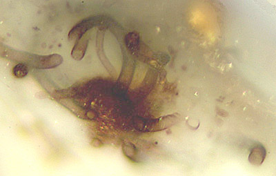
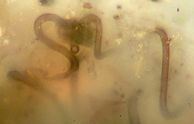
Fig.2 (far left): Nematoplexus
lump with tubes of 8...15µm, cut off at the sample surface so that
no full turns of spirals are seen. Detail from Fig.1, other
orientation.
Width of Figs.2,3: 0.3mm.
Fig.3: Uncommonly small
Nematoplexus
lump with two spiralling tubes, 9µm and 12µm, another one cut off at
the sample surface (= old fracture face), seen as a circular
cross-section. Detail from Fig.1, other
orientation.
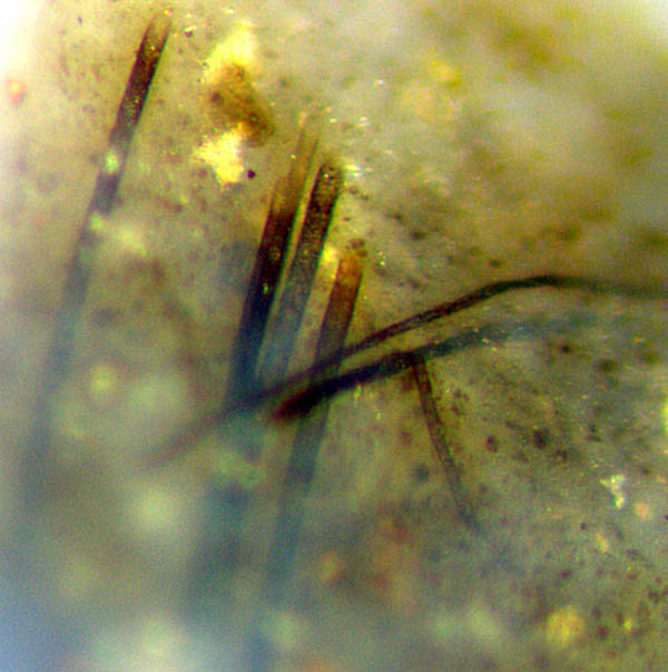
As one of the several peculiarities encountered in connection with Nematoplexus, tubes
appearing perfectly straight may unexpectedly be found near wound
tubes. The positions of Fig.1 and Fig.4, for example, are only a few
centimeters apart. While
the screw-like wound tubes known as spirals are produced by lumps
called branch-knots, the origin of the slightly curved tubes had been
unknown until evidence appeared which indicated that the bigger ones
are attached to
another kind of lumps. (See Rhynie
Chert News 71,
135.)
Hence, on can guess that the thinner slightly curved
tubes and the straight tubes come from some not yet seen type
of lump or knot.
The
idea that the tubes with distinctly different curvature might represent
different species is not favoured here since it would be improbable to
find them mostly near each other.
The conspicuous arrangement of straight tubes, parallel and in bundles,
requires an explanation, and so does the
presence of straight big tubes and curved narrow tubes close
together. The 5 parallel tubes arising from the
depth in Fig.4 meet the
sample surface where they end with elliptical sections. A 14µm-tube is
positioned behind a larger one, 2nd
from the left, so that its section is seen higher up in the picture. A
10µm-tube fitting nowhere incidentally ends
where a 12µm-tube is lying in the depth, providing the illusion of
rectangular branching.
Fig.4: Nematoplexus,
5 parallel straight tubes
14...21µm, slightly curved tubes 8 µm, 10µm, 12µm.
Picture taken at the raw sample surface, width 0.6mm,
same scale as Fig.1.
So it has appeared again that Nematoplexus
offers details which make
one wonder in which way they could possibly fit into a consistent
picture of this enigmatic organism. The problem is made even more
confusing by the non-spiralling
tubes with patterned walls resembling those of Nematothallus,
not shown here but often found along with
the spiralling smooth-walled tubes.
Comparison with several own finds of other nematophyte
species in the Rhynie chert have been of no much help.
All pictures have been taken from the raw
surface of one Rhynie chert sample of 0.28kg,
Rh9/86, found in 2003.
H.-J. Weiss
2019
[1] A.G. Lyon:
On the
fragmentary remains of an organism referable to the nematophytales,
from the
Rhynie chert, Nematoplexus
rhyniensis. Trans. Roy. Soc. Edinburgh
65(1961-62), 79-87, 2 tables.
[2] T.N. Taylor,
E.L. Taylor, M. Krings: Paleobotany,
Elsevier 2009.
[3]
www.abdn.ac.uk/rhynie/nemato.htm
 |
 |
136 |