Nematoplexus
-
thrice enigmatic
Nematoplexus
is properly listed
under the heading "Enigmatic Organisms" in [1] since this strange
creature consists essentially of tubes in several
varieties: ...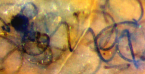
(1) nearly straight,
slightly curved, screw-like wound, or irregular,
(2) with smooth or patterned
walls and a wide range
of diameters,
(3) emerging from dark
lumps.
Most peculiar, several of these features may be found
combined in various ways: weakly curved thick and thin tubes side
by side, regular
screw-like spirals with smooth wall beside crooked tubes with spiral
wall patterning [2], small spirals beside big spirals, and more of such.
All this versatility is restricted by rules: The thread is always right-handed,
and its diameter is roughly equal to the pitch.
Along the tubes, their type does not change: Spirals remain screw-like,
diameter mostly below 16µm, and tubes with about 20µm never form
spirals.
These rules are to be considered preliminary since they are based on the
few recovered samples.
The inner workings of the
dark lumps, the
so-called branch
knots supposed to produce the
tubes, is an enigma in itself. Their
sizes vary between about 0.04mm and 0.25mm.
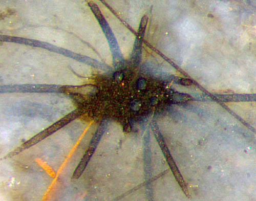
Fig.1 (right): Nematoplexus,
usual aspect with screw-like wound tubes and dark lump. Width of
Figs.1,2: 0.7mm.
Fig.2 (left): Slightly curved big tubes and
very thin ones emerging from "branch
knot",
also two unrelated straight ones, found near spiralling Nematoplexus .
One would never think of assigning the two quite different structures
seen in Fig.1 and Fig.2 to one species. Nevertheless,
one can suspect
that these structures might be mutually related
in some hitherto unknown way, being different manifestations
of the same organism. This suspicion is based on the observation that
the big slightly curved tubes with their knots are never far away
from the more slender spirals with their knots, at distances
of
about 2cm or less. They are very rare fossils, and
if they represented
independent organisms, it would be highly improbable to find the
different forms
closely together in one small chert sample.
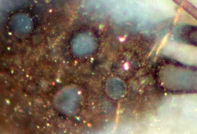
Fig.3 (right): Nematoplexus big
tubes emerging from the dark lump with obscure structure in
Fig.2. Width of the image 0.2mm.
"Branch knot" alludes to the notion that
"Branching of the tubes occurs in ...very tightly coiled knots of tubes
showing repeated and closely spaced branching" [3]. This is not
substantiated by the samples available here. In Figs.2,3 it is seen
that the tubes are
club-like where they emerge from the dark lump and
that there is no indication
of branching. The club-like shape is
also
obvious from the cross-sections of tubes cut near the lump, with
diameters up to 34µm near the lump in Fig.3 but 17...20µm farther out
in Fig.2. Thus it seems that
the surface of the lump, although not well defined, serves as a
reference site for the tubes. (See also Rhynie
Chert News 134.)
The
inclined cut on the right in Fig.3 looks as if the tube simply sticks
to the
surface of the lump.
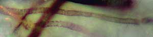
Fig.4 (left): Nematoplexus narrow
tubes (5µm) with patterned wall, detail from Fig.2, turned 90° to the right. Width 0.15mm, same scale as Fig.3.
Another remarkable feature in
Fig.2 is the narrow
filaments attached to the lump. Two of them are
faintly seen where an unrelated
straight narrow tube (8µm) crosses a cut-off big
tube. The irregular mixed annular and spiral pattern as known from tube
walls in the type specimen [2] is
seen here on the walls of the narrow tubes
of 5µm, with spacings of 2...3µm between the
dimly seen structure elements (Fig.4). All other nematophyte
tubes in this sample seem to have got smooth walls.
The
apparently radially extending big tubes in Fig.2 are really slightly
curved towards the observer, which is out of the picture plane, and accordingly are
cut off. 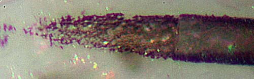 The
granular aspect of the big tubes in Fig.2 is possibly due to microbial
deposits or to decay products of the wall substance, as suggested by
Fig.5, which shows the tube bent out of the picture plane near the
lower edge of Fig.2. The outside of the tube enclosed in chalzedony is
seen
on the right in Fig.5. A clean slanting cut
would have produced a
parabolic boundary of the tube wall towards the left. Instead, a crack
had run across the wall and fragments must have fallen off. Farther
left, the rear wall with its probably
insignificant
random pattern is exposed.
The
granular aspect of the big tubes in Fig.2 is possibly due to microbial
deposits or to decay products of the wall substance, as suggested by
Fig.5, which shows the tube bent out of the picture plane near the
lower edge of Fig.2. The outside of the tube enclosed in chalzedony is
seen
on the right in Fig.5. A clean slanting cut
would have produced a
parabolic boundary of the tube wall towards the left. Instead, a crack
had run across the wall and fragments must have fallen off. Farther
left, the rear wall with its probably
insignificant
random pattern is exposed.
Fig.5: Slanting cut of a big tube in
Fig.2 with a probably irrelevant random pattern on the rear
wall. Width of the image 0.1mm.
From the samples available here it can be
concluded that Nematoplexus is
more complex than a mere meshwork of "spirally coiled
tubular cells" [3] and that it is not confined to the parameter region
stated in [2] for the type specimen of Nematoplexus rhyniensis.
This might be due to an extremely wide variability of the species or to
quite another cause: Nematoplexus might
be a symbiotic organism composed of several
partners contributing tubes of various sizes and
curvatures, with one or other of them occasionally dominating for
reasons unknown, forming shapes as in Figs.1,2.
More surprises can be expected from new finds.
All pictures have been taken from one sample labelled Rh15/79,
obtained from Barron in
2014, cut into 4 parts. Those with higher magnification, Figs.3-5, have
been taken from Part
1 by Gerd
Schmahl.in 2018.
H.-J. Weiss 2018
[1] T.N. Taylor,
E.L. Taylor, M. Krings: Paleobotany,
Elsevier 2009.
[2] A.G.
Lyon:
On the
fragmentary remains of an organism referable to the nematophytales,
from the Rhynie chert, Nematoplexus
rhyniensis.
Trans. Roy. Soc. Edinburgh
65(1961-62), 79-87, 2 tables.
[3]
www.abdn.ac.uk/rhynie/nemato.htm
 |
 |
135 |