Nematoplexus
surprisingly polymorphic
 The enigmatic organism Nematoplexus
has been known since 1961 as an assemblage of screw-like wound tubes,
diameters 7-10µm, emerging from dark lumps named "branch knots", in one
sample of Rhynie chert [1], then discovered as a smaller patch of such
tubes in a separate stratum of the Rhynie chert called
Windyfield chert, published in
2003 [2]. Also in 2003, samples were
found with similar-shaped but larger tubes, 10-15µm, also with slightly curved or straight ones near
the wound ones.
The enigmatic organism Nematoplexus
has been known since 1961 as an assemblage of screw-like wound tubes,
diameters 7-10µm, emerging from dark lumps named "branch knots", in one
sample of Rhynie chert [1], then discovered as a smaller patch of such
tubes in a separate stratum of the Rhynie chert called
Windyfield chert, published in
2003 [2]. Also in 2003, samples were
found with similar-shaped but larger tubes, 10-15µm, also with slightly curved or straight ones near
the wound ones.
What seemed to be a different species, big tubes
of about 20µm radially grown from a big dark lump, had first been seen
in a sample found in 2014 and described in Rhynie
Chert News 71.
Then, this combination of big tubes and big lump
had unexpectedly been seen in a second sample, and shown,
among other lumps, in Rhynie
Chert News 126.
The results of closer inspection of this same sample are presented
here.
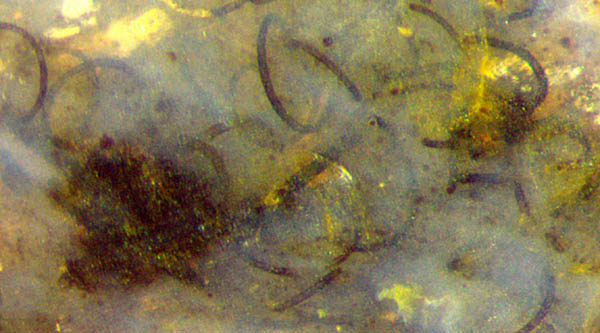
Fig.1: Nematoplexus
tubes and
"knots" on the raw surface of a small sample of 30g. Picture
width 1mm.
Closer inspection of Fig.1 suggests that the
"normal"
wound tubes are not
related to the big lump
which serves, although inconspicuously in the
present case,
as a center of much bigger tubes only. This is
revealed by stacks of pictures taken and combined into more distinct
images by Gerd
Schmahl:
see Figs.2,3.
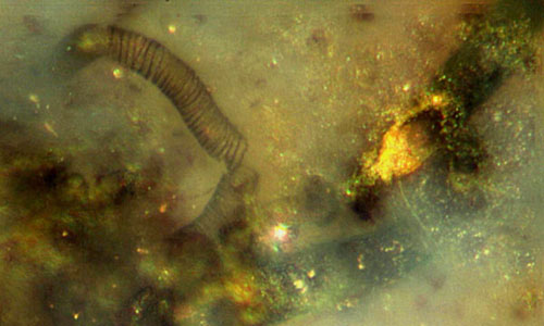
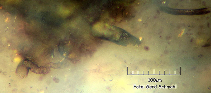
Fig.2 (left):
Right part of the big lump in Fig.1
with hollow big tube (right) and patterned
tube (left).
Picture width 0.25mm.
Fig.3 (right),
same scale as Fig.2: Lower part of the big lump in Fig.1,
slightly turned to the left.
Picture width 0.4mm.
Most conspicuous in Fig.2 is the inclined cut of
the hollow big tube with yellow deposit,
23µm outer diameter. Another tube of about 30µm is vaguely seen as a
broad dark strip in the depth below, more clearly seen on another
picture of the stack.
A stub of a big tube of 30µm is seen in
Fig.3. It seems to begin on a kind of surface, quite similar to a
same-size tube apparently attached to a big lump in Rhynie
Chert
News 135.
A curved 10µm-tube in Fig.3 above right represents the "normal" tubes
making the "Nematoplexus-Aspekt"
with the "normal" lumps
traditionally called branch-knots. The small lump in Fig.1 is probably
one of such.
The big lump in Fig.1 has got something in
common with other big lumps
shown in Rhynie
Chert
News 71, 126, 135:
It is the presence of big tubes (like those hidden in Fig.1 and made
visible in Figs.2,3) and the absence of adhearing
"normal"
Nematoplexus
tubes. This distinguishes them clearly from the
so-called "branch
knots", and since no branching has been seen, they may simply be called
"lumps with big tubes".
Also remarkable are the tubes with patterned wall. The
broken 17µm-tube in Fig.2
with irregular annular or spiral pattern appears surprisingly
similar to the tubes of Nematothallus
and also to the tracheids of vascular plants. 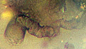
Fig.4:
Irregular-shaped tube with patterned wall and
variable diameter, detail of Fig.3 below left.
Picture width 0.1mm.
The unusual structure in Fig.4 is particularly interesting:
It is an irregular-shaped tube with patterned wall
and variable diameter: 13µm in the depth
and 23µm at the sample surface. Possibly
it had been hidden within the dark lump and is seen here only because
of
partial decay of the lump. It seems to turn into a regular
tube with patterned wall
while emerging from the lump on the left.
The slightly
curved or straight tubes mentioned
above have been found in this small sample, too: Figs.5,6.
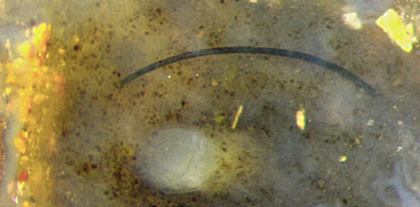
Fig.5 (left), same scale as Fig.1: Slightly bent Nematoplexus tube
(about 14µm) and charophyte branch section (0.15mm).
Picture width 1mm.
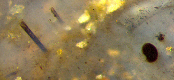
Fig.6 (right),
same scale as Fig.1:
Straight Nematoplexus tubes
(20µm, 31µm) and charophyte parts.Picture width 1.2mm.
As another unexplained phenomenon,
apparently parallel straight Nematoplexus tubes
with their origin hidden in the depth of the sample have been seen
before: Rhynie
Chert
News 136.
The tubes in Fig.6 have grown right through the
branches of a charophyte
alga, probably after the latter had been partially
decayed, except for the well-preserved dark capsule
tentatively interpreted as an oogonium.
For more details of this quite uncommon charophyte discovered in 2015
and found together with Nematoplexus
in the presently considered chert sample see Rhynie Chert
News 90.
Disregarding the not yet understood details
(and the tubes with patterned walls),
the
sparse fossil evidence justifies some
conclusions to be drawn:
- The screw-like wound tubes have got
diameters
of mostly 7-15µm and are associated with their kind of lumps
known as "branch
knots".
- The narrow weakly curved or straight
tubes have got diameters of mostly
7-15µm. Their related lumps have not yet been seen.
- The big weakly curved
or straight tubes have got diameters of
mostly 17-30µm and are associated with
the hitherto
unknown "lumps with big tubes".
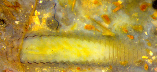
Fig.7: Detached Nematoplexus
tube fragment, 12µm, of non-typical shape;
crustacean Castracollis,
main body part seen from above.
Picture width 2.8mm.
Disregarding separate tube fragments
which do not fit into the above types, like the one in Fig.7,
the following can be stated:
No two of these three types of tubes have been seen transforming among
themselves or adhering to the same lump. In
particular, the winding tubes of 7-15µm are never seen emerging from
the "lumps with big tubes",
and the big tubes are never seen attached
to the "branch knots".
The subject is made even more confusing
by tubes with patterned walls and a wide range
of diameters,
known to arise from branch
knots. They can also arise from "lumps
with big tubes", as shown
here for the
first time.
With these observational facts one might be tempted to
assume that
there were two or more separate Nematoplexus
species
if there were not the unexpected facts that the weakly curved tubes are
always near or between the
screw-like wound tubes and that the big and narrow weakly
curved or straight tubes are mostly found together in one sample.
If the tubes represented separate species,
it would be highly improbable to find tubes of two or three different
types nearly always together in one sample. Since they are
found together in
small chert samples like the present one, there must be a reason.
Possible reasons are hard to imagine, and any one would be highly
unusual: some kind of symbiotic relation between two nematophyte
species, or Nematoplexus with
an ability to bring forth various forms, for whichever reason. Anyway, Nematoplexus can
be expected to provide more surprises.
All pictures have been taken from the natural surface of one chert sample of 30g,
Rh6/102, found by Sieglinde Weiss in 2003.
H.-J. Weiss 2019
[1] A.G. Lyon:
On the fragmentary remains of an organism referable to the
nematophytales,
from the
Rhynie chert, Nematoplexus
rhyniensis. Trans. Roy. Soc. Edinburgh
65(1961-62), 79-87, 2 tables.
[2] S.R.
Fayers, N.H. Trewin: A
review of the palaeoinvironments and biota of the Windyfield chert.
Trans. Roy. Soc. Edinburgh Earth Sci. 94(2004 for 2003), 325-39.
[3]
www.abdn.ac.uk/rhynie/nemato.htm
 |
 |
137 |